Our official English website, www.x-mol.net, welcomes your feedback! (Note: you will need to create a separate account there.)
Comparison of SB-SDS and other decellularization methods for the acellular nerve graft: Biological evaluation and nerve repair in vitro and in vivo.
SYNAPSE ( IF 2.3 ) Pub Date : 2019-11-09 , DOI: 10.1002/syn.22143 Li-Wei Han 1 , Gang Xu 2, 3 , Mei-Yu Guo 2, 3 , Yv-Ang Chang 2, 3 , Yu Zhang 2, 3 , Yan-Tao Zhao 1 , Zhong-Hai Li 2, 3
SYNAPSE ( IF 2.3 ) Pub Date : 2019-11-09 , DOI: 10.1002/syn.22143 Li-Wei Han 1 , Gang Xu 2, 3 , Mei-Yu Guo 2, 3 , Yv-Ang Chang 2, 3 , Yu Zhang 2, 3 , Yan-Tao Zhao 1 , Zhong-Hai Li 2, 3
Affiliation
|
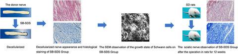
We aimed to compare the performance of acellular nerves prepared by different decellularization methods, screening out the optimal decellularization protocol, repairing the sciatic nerve defects in rats by the allogeneic transplantation, and evaluating the effect of regenerative nerve on the function reconstruction. The Sondell, SB-SDS, TnBP, and the high/low permeation methods were used to decellularize donor nerves. Nerves without any treatment were as the control group. The histological results were evaluated by HE staining and toluidine blue (TB) staining. The proliferation activity of L929 cells was detected by CCK-8 assay. The adhesion of Schwann cells was observed and quantified by SEM. Balb/c mice were used to evaluate the cellular and humoral immunogenicity of the nerve scaffolds. The rat sciatic nerve defect model was applied to observe the repair effect of acellular nerve scaffold in vivo. To SB-SDS group, it remained the original state of the nerves, with no observed nucleus and axons, the neurotoxicity grade detected by CCK-8 being almost 0, and it kept the largest number of Schwann cells adhered to the acellular nerve and the better morphology. Further, it showed that the selected SB-SDS rats acellular nerve scaffold could promote the nerve repair of the rats by HE staining and TB staining. We could conclude that the acellular nerve matrix prepared by the SB-SDS method effectively removes the cellular components in the nerve tissue and retains the main components of the extracellular matrix of the nerve tissue, whose rats decellularized nerve scaffold could promote the sciatic nerve repair better.
中文翻译:

SB-SDS 与其他脱细胞神经移植方法的比较:体外和体内生物学评价和神经修复。
我们旨在比较不同脱细胞方法制备的脱细胞神经的性能,筛选最佳脱细胞方案,通过同种异体移植修复大鼠坐骨神经缺损,并评估再生神经对功能重建的影响。Sondell、SB-SDS、TnBP 和高/低渗透方法用于使供体神经脱细胞。未进行任何处理的神经作为对照组。通过HE染色和甲苯胺蓝(TB)染色评估组织学结果。CCK-8法检测L929细胞增殖活性。通过扫描电镜观察和量化雪旺氏细胞的粘附。Balb/c 小鼠用于评估神经支架的细胞和体液免疫原性。应用大鼠坐骨神经缺损模型观察脱细胞神经支架在体内的修复效果。SB-SDS组保持神经原始状态,未观察到细胞核和轴突,CCK-8检测的神经毒性等级几乎为0,并保留了最大数量的雪旺细胞粘附于脱细胞神经和更好的形态。HE染色和TB染色表明,所选择的SB-SDS大鼠脱细胞神经支架能够促进大鼠的神经修复。可以得出结论,SB-SDS法制备的脱细胞神经基质能有效去除神经组织中的细胞成分,保留神经组织细胞外基质的主要成分,大鼠脱细胞神经支架能更好地促进坐骨神经修复。 .
更新日期:2020-03-27
中文翻译:
SB-SDS 与其他脱细胞神经移植方法的比较:体外和体内生物学评价和神经修复。
我们旨在比较不同脱细胞方法制备的脱细胞神经的性能,筛选最佳脱细胞方案,通过同种异体移植修复大鼠坐骨神经缺损,并评估再生神经对功能重建的影响。Sondell、SB-SDS、TnBP 和高/低渗透方法用于使供体神经脱细胞。未进行任何处理的神经作为对照组。通过HE染色和甲苯胺蓝(TB)染色评估组织学结果。CCK-8法检测L929细胞增殖活性。通过扫描电镜观察和量化雪旺氏细胞的粘附。Balb/c 小鼠用于评估神经支架的细胞和体液免疫原性。应用大鼠坐骨神经缺损模型观察脱细胞神经支架在体内的修复效果。SB-SDS组保持神经原始状态,未观察到细胞核和轴突,CCK-8检测的神经毒性等级几乎为0,并保留了最大数量的雪旺细胞粘附于脱细胞神经和更好的形态。HE染色和TB染色表明,所选择的SB-SDS大鼠脱细胞神经支架能够促进大鼠的神经修复。可以得出结论,SB-SDS法制备的脱细胞神经基质能有效去除神经组织中的细胞成分,保留神经组织细胞外基质的主要成分,大鼠脱细胞神经支架能更好地促进坐骨神经修复。 .


 京公网安备 11010802027423号