当前位置:
X-MOL 学术
›
Dev. Growth Differ.
›
论文详情
Our official English website, www.x-mol.net, welcomes your feedback! (Note: you will need to create a separate account there.)
User-friendly cell patterning methods using a polydimethylsiloxane mold with microchannels.
Development, Growth & Differentiation ( IF 2.5 ) Pub Date : 2019-11-21 , DOI: 10.1111/dgd.12637 Shun-Ichi Funano 1 , Nobuyuki Tanaka 1 , Yo Tanaka 1
Development, Growth & Differentiation ( IF 2.5 ) Pub Date : 2019-11-21 , DOI: 10.1111/dgd.12637 Shun-Ichi Funano 1 , Nobuyuki Tanaka 1 , Yo Tanaka 1
Affiliation
|
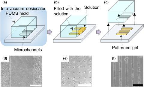
Techniques for partitioning cell adhesion are useful tools in biological and medical experiments. However, conventional cell patterning methods require special apparatus, special materials or high-level skills. Therefore, we have developed a new cell patterning methodology which can be easily carried out in biological laboratories. Non-cell adhesive material including hydrogel or gas patterns to restrict cell adhesion on a culture dish or glass substrates can be constructed by exploiting a polydimethylsiloxane (PDMS) mold with microchannels. The PDMS molds suck non-adhesive materials into microchannels from the inlet of the microchannels and the materials are immobilized onto the substrates with a desired pattern. High resolution under a few micrometers and long-term stability can be realized. This method has been used for analysis of stem cells, muscle cells, neuron development and other cells in collaboration with many biological researchers. Several examples to use this technique are introduced in this review.
中文翻译:

使用带有微通道的聚二甲基硅氧烷模具的用户友好型电池图案化方法。
分配细胞粘附的技术是生物学和医学实验中的有用工具。然而,常规的单元图案化方法需要特殊的设备,特殊的材料或高水平的技能。因此,我们开发了一种新的细胞模式方法,可以在生物实验室中轻松进行。可以通过利用具有微通道的聚二甲基硅氧烷(PDMS)模具来构建包括水凝胶或气体图案以限制细胞在培养皿或玻璃基板上粘附的非细胞粘附材料。PDMS模具将非粘性材料从微通道的入口吸入微通道,然后将材料固定在具有所需图案的基材上。几微米下即可实现高分辨率,并具有长期稳定性。此方法已用于分析干细胞,肌肉细胞,神经元发育和其他细胞与许多生物学研究人员合作。在这篇评论中介绍了使用这种技术的几个例子。
更新日期:2019-11-21
中文翻译:
使用带有微通道的聚二甲基硅氧烷模具的用户友好型电池图案化方法。
分配细胞粘附的技术是生物学和医学实验中的有用工具。然而,常规的单元图案化方法需要特殊的设备,特殊的材料或高水平的技能。因此,我们开发了一种新的细胞模式方法,可以在生物实验室中轻松进行。可以通过利用具有微通道的聚二甲基硅氧烷(PDMS)模具来构建包括水凝胶或气体图案以限制细胞在培养皿或玻璃基板上粘附的非细胞粘附材料。PDMS模具将非粘性材料从微通道的入口吸入微通道,然后将材料固定在具有所需图案的基材上。几微米下即可实现高分辨率,并具有长期稳定性。此方法已用于分析干细胞,肌肉细胞,神经元发育和其他细胞与许多生物学研究人员合作。在这篇评论中介绍了使用这种技术的几个例子。


 京公网安备 11010802027423号