当前位置:
X-MOL 学术
›
Adv. Mater.
›
论文详情
Our official English website, www.x-mol.net, welcomes your feedback! (Note: you will need to create a separate account there.)
Aqueous Two‐Phase Emulsion Bioink‐Enabled 3D Bioprinting of Porous Hydrogels
Advanced Materials ( IF 29.4 ) Pub Date : 2018-10-21 , DOI: 10.1002/adma.201805460 Guo-Liang Ying 1, 2 , Nan Jiang 3 , Sushila Maharjan 1, 4 , Yi-Xia Yin 1, 5 , Rong-Rong Chai 1 , Xia Cao 1 , Jing-Zhou Yang 1, 6 , Amir K Miri 1 , Shabir Hassan 1 , Yu Shrike Zhang 1
Advanced Materials ( IF 29.4 ) Pub Date : 2018-10-21 , DOI: 10.1002/adma.201805460 Guo-Liang Ying 1, 2 , Nan Jiang 3 , Sushila Maharjan 1, 4 , Yi-Xia Yin 1, 5 , Rong-Rong Chai 1 , Xia Cao 1 , Jing-Zhou Yang 1, 6 , Amir K Miri 1 , Shabir Hassan 1 , Yu Shrike Zhang 1
Affiliation
|
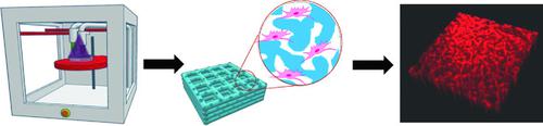
3D bioprinting technology provides programmable and customizable platforms to engineer cell‐laden constructs mimicking human tissues for a wide range of biomedical applications. However, the encapsulated cells are often restricted in spreading and proliferation by dense biomaterial networks from gelation of bioinks. Herein, a cell‐benign approach is reported to directly bioprint porous‐structured hydrogel constructs by using an aqueous two‐phase emulsion bioink. The bioink, which contains two immiscible aqueous phases of cell/gelatin methacryloyl (GelMA) mixture and poly(ethylene oxide) (PEO), is photocrosslinked to fabricate predesigned cell‐laden hydrogel constructs by extrusion bioprinting or digital micromirror device‐based stereolithographic bioprinting. The porous structure of the 3D‐bioprinted hydrogel construct is formed by subsequently removing the PEO phase from the photocrosslinked GelMA hydrogel. Three different cell types (human hepatocellular carcinoma cells, human umbilical vein endothelial cells, and NIH/3T3 mouse embryonic fibroblasts) within the 3D‐bioprinted porous hydrogel patterns show enhanced cell viability, spreading, and proliferation compared to the standard (i.e., nonporous) hydrogel constructs. The 3D bioprinting strategy is believed to provide a robust and versatile platform to engineer porous‐structured tissue constructs and their models for a variety of applications in tissue engineering, regenerative medicine, drug development, and personalized therapeutics.
中文翻译:

水性两相乳液生物墨水启用的多孔水凝胶 3D 生物打印
3D 生物打印技术提供了可编程和可定制的平台来设计模仿人体组织的充满细胞的结构,用于广泛的生物医学应用。然而,封装的细胞的扩散和增殖通常受到生物墨水凝胶化的致密生物材料网络的限制。本文报道了一种细胞良性方法,通过使用水性两相乳液生物墨水直接生物打印多孔结构水凝胶结构。该生物墨水含有细胞/明胶甲基丙烯酰基 (GelMA) 混合物和聚环氧乙烷 (PEO) 的两个不混溶水相,通过光交联,通过挤出生物打印或基于数字微镜设备的立体光刻生物打印来制造预先设计的充满细胞的水凝胶结构。随后从光交联的 GelMA 水凝胶中去除 PEO 相,形成 3D 生物打印水凝胶结构的多孔结构。与标准(即无孔)相比,3D生物打印的多孔水凝胶图案中的三种不同细胞类型(人肝细胞癌细胞、人脐静脉内皮细胞和NIH/3T3小鼠胚胎成纤维细胞)显示出增强的细胞活力、扩散和增殖能力水凝胶结构。3D生物打印策略被认为提供了一个强大且多功能的平台来设计多孔结构组织结构及其模型,用于组织工程、再生医学、药物开发和个性化治疗的各种应用。
更新日期:2018-10-21
中文翻译:
水性两相乳液生物墨水启用的多孔水凝胶 3D 生物打印
3D 生物打印技术提供了可编程和可定制的平台来设计模仿人体组织的充满细胞的结构,用于广泛的生物医学应用。然而,封装的细胞的扩散和增殖通常受到生物墨水凝胶化的致密生物材料网络的限制。本文报道了一种细胞良性方法,通过使用水性两相乳液生物墨水直接生物打印多孔结构水凝胶结构。该生物墨水含有细胞/明胶甲基丙烯酰基 (GelMA) 混合物和聚环氧乙烷 (PEO) 的两个不混溶水相,通过光交联,通过挤出生物打印或基于数字微镜设备的立体光刻生物打印来制造预先设计的充满细胞的水凝胶结构。随后从光交联的 GelMA 水凝胶中去除 PEO 相,形成 3D 生物打印水凝胶结构的多孔结构。与标准(即无孔)相比,3D生物打印的多孔水凝胶图案中的三种不同细胞类型(人肝细胞癌细胞、人脐静脉内皮细胞和NIH/3T3小鼠胚胎成纤维细胞)显示出增强的细胞活力、扩散和增殖能力水凝胶结构。3D生物打印策略被认为提供了一个强大且多功能的平台来设计多孔结构组织结构及其模型,用于组织工程、再生医学、药物开发和个性化治疗的各种应用。


 京公网安备 11010802027423号