Our official English website, www.x-mol.net, welcomes your feedback! (Note: you will need to create a separate account there.)
Distinct Iron Deposition Profiles of Liver Zones in Various Models with Iron Homeostasis Disorders
Advanced Science ( IF 15.1 ) Pub Date : 2018-10-12 , DOI: 10.1002/advs.201800866 Haoyang Song 1, 2 , Shuping Zhang 3 , Xia Sun 4 , Jing Liu 2 , Yakun Wu 2, 5 , Wenli Guo 6, 7 , Fudi Wang 8 , Xiaojuan Ou 9 , Min Cong 9 , Erhu Jin 4 , Wenyong Li 1 , Sijin Liu 2, 5
Advanced Science ( IF 15.1 ) Pub Date : 2018-10-12 , DOI: 10.1002/advs.201800866 Haoyang Song 1, 2 , Shuping Zhang 3 , Xia Sun 4 , Jing Liu 2 , Yakun Wu 2, 5 , Wenli Guo 6, 7 , Fudi Wang 8 , Xiaojuan Ou 9 , Min Cong 9 , Erhu Jin 4 , Wenyong Li 1 , Sijin Liu 2, 5
Affiliation
|
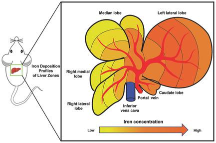
Determination of iron accumulation is crucial in diagnosing the occurrence and progression of many liver‐ and iron‐related diseases. Thus far, little is known about the profiles of iron deposition in different liver zones, particularly under conditions with disordered iron homeostasis. Here, uneven iron distribution in livers of patients with hereditary hemochromatosis (HH) is uncovered, showing the region with the highest iron concentration near the entrance site of the portal vein and hepatic artery in contrast to the sites with the lowest iron concentration close to the distal edge. Distinct iron distribution profiles are also found throughout liver zones in wild‐type mice and various mouse models with iron metabolism disorders, including hemochromatosis (Hfe−/−), iron deficiency, and inflammation. Of note, similar findings observed in HH patients are further demonstrated in Hfe−/− mice. Moreover, the zones with greater iron accumulation appear to be more sensitive to iron changes, e.g., there is iron increase upon iron overload and iron loss in response to iron deficiency. Mechanistic investigation manifests that these differential iron changes in liver zones are subjected to the regulation by the hepcidin–ferroportin axis. Additionally, the data corroborate the reliability of magnetic resonance imaging (MRI) in recognizing the differential iron deposition profiles among liver zones.
中文翻译:

具有铁稳态紊乱的各种模型中肝区的独特铁沉积特征
铁积累的测定对于诊断许多肝脏和铁相关疾病的发生和进展至关重要。迄今为止,人们对不同肝区铁沉积的情况知之甚少,特别是在铁稳态紊乱的情况下。这里揭示了遗传性血色素沉着病 (HH) 患者肝脏中铁分布不均匀的情况,显示靠近门静脉和肝动脉入口部位铁浓度最高的区域,与靠近门静脉和肝动脉入口部位铁浓度最低的部位形成鲜明对比。远端边缘。在野生型小鼠和各种患有铁代谢紊乱(包括血色素沉着症(Hfe −/− )、缺铁和炎症的小鼠模型)的肝区中也发现了不同的铁分布特征。值得注意的是,在 HH 患者中观察到的类似结果在Hfe −/−小鼠中得到了进一步证实。此外,铁积累较多的区域似乎对铁的变化更敏感,例如,铁过载时铁增加,铁缺乏时铁损失。机制研究表明,肝区铁的这些差异变化受到铁调素-铁转运蛋白轴的调节。此外,这些数据证实了磁共振成像(MRI)在识别肝区铁沉积差异方面的可靠性。
更新日期:2018-10-12
中文翻译:
具有铁稳态紊乱的各种模型中肝区的独特铁沉积特征
铁积累的测定对于诊断许多肝脏和铁相关疾病的发生和进展至关重要。迄今为止,人们对不同肝区铁沉积的情况知之甚少,特别是在铁稳态紊乱的情况下。这里揭示了遗传性血色素沉着病 (HH) 患者肝脏中铁分布不均匀的情况,显示靠近门静脉和肝动脉入口部位铁浓度最高的区域,与靠近门静脉和肝动脉入口部位铁浓度最低的部位形成鲜明对比。远端边缘。在野生型小鼠和各种患有铁代谢紊乱(包括血色素沉着症(Hfe −/− )、缺铁和炎症的小鼠模型)的肝区中也发现了不同的铁分布特征。值得注意的是,在 HH 患者中观察到的类似结果在Hfe −/−小鼠中得到了进一步证实。此外,铁积累较多的区域似乎对铁的变化更敏感,例如,铁过载时铁增加,铁缺乏时铁损失。机制研究表明,肝区铁的这些差异变化受到铁调素-铁转运蛋白轴的调节。此外,这些数据证实了磁共振成像(MRI)在识别肝区铁沉积差异方面的可靠性。


 京公网安备 11010802027423号