Theranostics ( IF 12.4 ) Pub Date : 2018-09-09 , DOI: 10.7150/thno.24252 Ayman El-Sayed , Wendy Bernhard , Kris Barreto , Carolina Gonzalez , Wayne Hill , Landon Pastushok , Humphrey Fonge , C. Ronald Geyer
|
In vivo imaging is influenced by the half-life, tissue penetration, biodistribution, and affinity of the imaging probe. Immunoglobulin G (IgG) is composed of discrete domains with known functions, providing a template for engineering antibody fragments with desired imaging properties. Here, we engineered antibody-based imaging probes, consisting of different combinations of antibody domains, labeled them with the near-infrared fluorescent dye IRDye800CW, and evaluated their in vivo imaging properties. Antibody-based imaging probes were based on an anti-HER3 antigen binding fragment (Fab) isolated using phage display.
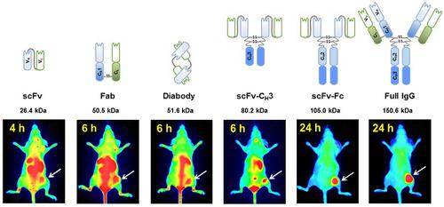
Methods: We constructed six anti-HER3 antibody-based imaging probes: a single chain variable fragment (scFv), Fab, diabody, scFv-CH3, scFv-Fc, and IgG. IRDye800CW-labeled, antibody-based probes were injected into nude mice bearing FaDu xenografts and their distribution to the xenograft, liver, and kidneys was evaluated.
Results: These imaging probes bound to recombinant HER3 and to the HER3-positive cell line, FaDu. Small antibody fragments with molecular weight <60 kDa (scFv, diabody, and Fab) accumulated rapidly in the xenograft (maximum accumulation between 2-4 h post injection (hpi)) and cleared primarily through the kidneys. scFv-CH3 (80 kDa) had fast clearance and peaked in the xenograft between 2-3 hpi and cleared from xenograft in a rate comparable to Fab and diabody. IgG and scFv-Fc persisted in the xenografts for up to 72 hpi and distributed mainly to the xenograft and liver. The highest xenograft fluorescence signals were observed with IgG and scFv-Fc imaging probes and persisted for 2-3 days.
Conclusion: These results highlight the utility of using antibody fragments to optimize clearance, tumor labeling, and biodistribution properties for developing anti-HER3 probes for image-guided surgery or PET imaging.
Keywords: antibody fragments, near-infrared fluorescence imaging, HER3, ErbB3, IRDye800CW
中文翻译:
HER3阳性癌症异种移植物近红外荧光成像抗体片段特性的评估
体内成像受成像探针的半衰期,组织渗透,生物分布和亲和力的影响。免疫球蛋白G(IgG)由具有已知功能的离散域组成,可为具有所需成像特性的工程抗体片段提供模板。在这里,我们设计了由抗体域的不同组合组成的基于抗体的成像探针,并用近红外荧光染料IRDye800CW对其进行了标记,并评估了它们的体内成像特性。基于抗体的成像探针基于使用噬菌体展示分离的抗HER3抗原结合片段(Fab)。
方法:我们构建了六种基于抗HER3抗体的成像探针:单链可变片段(scFv),Fab,双抗体,scFv-C H 3,scFv-Fc和IgG。将IRDye800CW标记的,基于抗体的探针注入带有FaDu异种移植物的裸鼠中,并评估它们在异种移植物,肝脏和肾脏中的分布。
结果:这些成像探针与重组HER3和HER3阳性细胞系FaDu结合。分子量小于60 kDa的小抗体片段(scFv,双抗体和Fab)在异种移植物中迅速积累(注射后2-4小时(hpi)之间最大积累)并主要通过肾脏清除。scFv-C H 3(80 kDa)具有快速清除能力,并在2-3 hpi之间在异种移植物中达到峰值,并以与Fab和双抗体相当的速率从异种移植物中清除。IgG和scFv-Fc在异种移植物中持续长达72 hpi,并主要分布于异种移植物和肝脏。用IgG和scFv-Fc成像探针观察到最高的异种移植荧光信号,并持续2-3天。
结论:这些结果突出了使用抗体片段优化清除率,肿瘤标记和生物分布特性的实用性,以开发用于图像引导手术或PET成像的抗HER3探针。
关键词:抗体片段,近红外荧光成像,HER3,ErbB3,IRDye800CW


 京公网安备 11010802027423号