Theranostics ( IF 12.4 ) Pub Date : 2018-09-09 , DOI: 10.7150/thno.24747 Conner Adams , Paolo Bazzigaluppi , Tina L. Beckett , Jossana Bishay , Iliya Weisspapir , Adrienne Dorr , James R. Mester , Joe Steinman , Lydiane Hirschler , Jan M. Warnking , Emmanuel L. Barbier , JoAnne McLaurin , John G. Sled , Bojana Stefanovic
|
Traumatic brain injury (TBI) research has focused on moderate to severe injuries as their outcomes are significantly worse than those of a mild TBI (mTBI). However, recent epidemiological evidence has indicated that a series of even mild TBIs greatly increases the risk of neurodegenerative and psychiatric disorders. Neuropathological studies of repeated TBI have identified changes in neuronal ionic concentrations, axonal injury, and cytoskeletal damage as important determinants of later life neurological and mood compromise; yet, there is a paucity of data on the contribution of neurogliovascular dysfunction to the progression of repeated TBI and alterations of brain function in the intervening period.
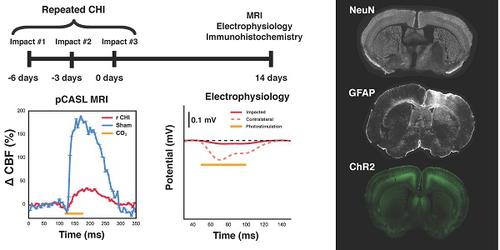
Methods: Here, we established a mouse model of repeated TBI induced via three electromagnetically actuated impacts delivered to the intact skull at three-day intervals and determined the long-term deficits in neurogliovascular functioning in Thy1-ChR2 mice. Two weeks post the third impact, cerebral blood flow and cerebrovascular reactivity were measured with arterial spin labelling magnetic resonance imaging. Neuronal function was investigated through bilateral intracranial electrophysiological responses to optogenetic photostimulation. Vascular density of the site of impacts was measured with in vivo two photon fluorescence microscopy. Pathological analysis of neuronal survival and astrogliosis was performed via NeuN and GFAP immunofluorescence.
Results: Cerebral blood flow and cerebrovascular reactivity were decreased by 50±16% and 70±20%, respectively, in the TBI cohort relative to sham-treated animals. Concomitantly, electrophysiological recordings revealed a 97±1% attenuation in peri-contusional neuronal reactivity relative to sham. Peri-contusional vascular volume was increased by 33±2% relative to sham-treated mice. Pathological analysis of the peri-contusional cortex demonstrated astrogliosis, but no changes in neuronal survival.
Conclusion: This work provides the first in-situ characterization of the long-term deficits of the neurogliovascular unit following repeated TBI. The findings will help guide the development of diagnostic markers as well as therapeutics targeting neurogliovascular dysfunction.
Keywords: repeated TBI, neurovascular, optogenetics, closed head injury
中文翻译:
重复性脑外伤模型中的神经胶质血管功能异常
创伤性脑损伤(TBI)的研究集中于中度至重度损伤,因为它们的结果明显比轻度TBI(mTBI)的结果差。但是,最近的流行病学证据表明,一系列甚至轻度的TBI也大大增加了神经退行性疾病和精神疾病的风险。反复进行TBI的神经病理学研究已确定神经元离子浓度,轴突损伤和细胞骨架损伤的改变是以后神经系统和情绪受损的重要决定因素。然而,在此期间,关于神经胶质血管功能障碍对反复TBI进展和脑功能改变的贡献还很少。
方法:在这里,我们建立了一个小鼠重复性TBI模型,该模型是通过三天间隔将三个电磁致动的冲击传递给完整的颅骨而诱发的,并确定了Thy1-ChR2小鼠的神经胶质血管功能的长期缺陷。第三次撞击后两周,通过动脉自旋标记磁共振成像测量脑血流量和脑血管反应性。通过对光遗传学光刺激的双侧颅内电生理反应研究了神经元功能。用体内两个光子荧光显微镜测量了撞击部位的血管密度。通过NeuN和GFAP免疫荧光进行神经元存活和星形胶质细胞变性的病理分析。
结果:与假手术组相比,TBI组的脑血流量和脑血管反应性分别降低了50±16%和70±20%。同时,电生理记录显示,与假手术相比,挫伤周围神经元反应性降低了97±1%。相对于假治疗的小鼠,挫伤周围的血管体积增加了33±2%。挫伤周围皮质的病理分析显示星形胶质增生,但神经元存活无变化。
结论:这项工作提供了反复TBI后神经胶质血管单位长期缺陷的第一个原位表征。这些发现将有助于指导诊断标记物以及针对神经胶质血管功能障碍的治疗方法的开发。
关键词:反复TBI,神经血管,光遗传学,闭合性颅脑损伤


 京公网安备 11010802027423号