Our official English website, www.x-mol.net, welcomes your feedback! (Note: you will need to create a separate account there.)
Observing and tracking single small ribosomal subunits in vivo
Methods ( IF 4.8 ) Pub Date : 2019-01-01 , DOI: 10.1016/j.ymeth.2018.09.001 Lisa Landvogt , Jan Andreas Ruland , Christian Montellese , Jan Peter Siebrasse , Ulrike Kutay , Ulrich Kubitscheck
Methods ( IF 4.8 ) Pub Date : 2019-01-01 , DOI: 10.1016/j.ymeth.2018.09.001 Lisa Landvogt , Jan Andreas Ruland , Christian Montellese , Jan Peter Siebrasse , Ulrike Kutay , Ulrich Kubitscheck
|
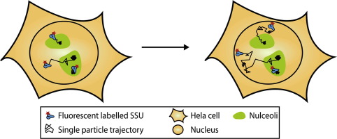
Ribosomes are formed of a small and a large subunit (SSU/LSU), both consisting of rRNA and a plethora of accessory proteins. While biochemical and genetic studies identified most of the involved proteins and deciphered the ribosomal synthesis steps, our knowledge of the molecular dynamics of the different ribosomal subunits and also of the kinetics of their intracellular trafficking is still limited. Adopting a labelling strategy initially used to study mRNA export we were able to fluorescently stain the SSU in vivo. We chose DIM2/PNO1 (Defective In DNA Methylation 2/Partner of NOb1) as labelling target and created a stable cell line carrying an inducible SNAP-DIM2 fusion protein. After bulk labelling with a green fluorescent dye combined with very sparse labelling with a red fluorescent dye the nucleoli and single SSU could be visualized simultaneously in the green and red channel, respectively. We used single molecule microscopy to track single SSU in the nucleolus and nucleoplasm. Resulting trajectory data were analyzed by jump-distance analysis and the variational Bayes single-particle tracking approach. Both methods allowed identifying the number of diffusive states and the corresponding diffusion coefficients. For both nucleoli and nucleoplasm we could identify mobile (D = 2.3-2.8 µm2/s), retarded (D = 0.18-0.31 µm2/s) and immobilized (D = 0.04-0.05 µm2/s) SSU fractions and, as expected, the size of the fractions differed in the two compartments. While the fast mobility fraction matches perfectly the expected nuclear mobility of the SSU (D = 2.45 µm2/s), we were surprised to find a substantial fraction (33%) of immobile SSU in the nucleoplasm, something not observed for inert control molecules.
中文翻译:

在体内观察和跟踪单个小核糖体亚基
核糖体由小亚基和大亚基 (SSU/LSU) 组成,均由 rRNA 和大量辅助蛋白组成。虽然生化和遗传研究确定了大部分涉及的蛋白质并破译了核糖体合成步骤,但我们对不同核糖体亚基的分子动力学及其细胞内运输动力学的了解仍然有限。采用最初用于研究 mRNA 输出的标记策略,我们能够在体内对 SSU 进行荧光染色。我们选择 DIM2/PNO1(DNA 甲基化缺陷 2/NOb1 的伙伴)作为标记目标,并创建了一个携带可诱导 SNAP-DIM2 融合蛋白的稳定细胞系。使用绿色荧光染料进行批量标记并使用红色荧光染料进行非常稀疏的标记后,可以分别在绿色和红色通道中同时观察到核仁和单个 SSU。我们使用单分子显微镜来跟踪核仁和核质中的单个 SSU。所得轨迹数据通过跳跃距离分析和变分贝叶斯单粒子跟踪方法进行分析。这两种方法都允许识别扩散状态的数量和相应的扩散系数。对于核仁和核质,我们可以识别移动 (D = 2.3-2.8 µm2/s)、延迟 (D = 0.18-0.31 µm2/s) 和固定 (D = 0.04-0.05 µm2/s) SSU 分数,正如预期的那样,两个隔室中馏分的大小不同。
更新日期:2019-01-01
中文翻译:
在体内观察和跟踪单个小核糖体亚基
核糖体由小亚基和大亚基 (SSU/LSU) 组成,均由 rRNA 和大量辅助蛋白组成。虽然生化和遗传研究确定了大部分涉及的蛋白质并破译了核糖体合成步骤,但我们对不同核糖体亚基的分子动力学及其细胞内运输动力学的了解仍然有限。采用最初用于研究 mRNA 输出的标记策略,我们能够在体内对 SSU 进行荧光染色。我们选择 DIM2/PNO1(DNA 甲基化缺陷 2/NOb1 的伙伴)作为标记目标,并创建了一个携带可诱导 SNAP-DIM2 融合蛋白的稳定细胞系。使用绿色荧光染料进行批量标记并使用红色荧光染料进行非常稀疏的标记后,可以分别在绿色和红色通道中同时观察到核仁和单个 SSU。我们使用单分子显微镜来跟踪核仁和核质中的单个 SSU。所得轨迹数据通过跳跃距离分析和变分贝叶斯单粒子跟踪方法进行分析。这两种方法都允许识别扩散状态的数量和相应的扩散系数。对于核仁和核质,我们可以识别移动 (D = 2.3-2.8 µm2/s)、延迟 (D = 0.18-0.31 µm2/s) 和固定 (D = 0.04-0.05 µm2/s) SSU 分数,正如预期的那样,两个隔室中馏分的大小不同。


 京公网安备 11010802027423号