Acta Biomaterialia ( IF 9.7 ) Pub Date : 2018-08-30 , DOI: 10.1016/j.actbio.2018.08.034 R. Amberg , A. Elad , D. Rothamel , T. Fienitz , G. Szakacs , S. Heilmann , F. Witte
|
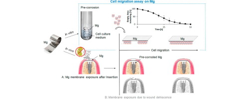
A novel regenerative approach to Guided Bone Regeneration (GBR) in dental surgery is based on the development of biodegradable and volume stable barrier membranes made of metallic magnesium. Currently used volume stable barrier membranes are made of titanium-reinforced PTFE or titanium-reinforced collagen membranes, both, however, are accompanied by a high incidence of wound dehiscence resulting in membrane exposure, which leads to an increased infection risk. An exposed membrane could also occur directly after insertion due to insufficient soft tissue coverage of the membrane. In both cases, fast wound margin regeneration is required. As a first step of soft-tissue regeneration, gingival fibroblasts need to migrate over the barrier membrane and close the dehiscent wound. Based on this aim, this study investigated the migration behaviour of human gingival fibroblasts on a magnesium surface. Major experimental challenges such as formation of hydrogen bubbles due to initial magnesium corrosion and non-transparent material surfaces have been addressed to allow cell adhesion and to follow cell migration. The designed scratch-based cell migration assay involved vital fluorescent cell staining on a pre-corroded magnesium membrane to simulate in vivo wound dehiscence. The assay has been used to compare cell migration on pre-corroded magnesium to titanium surfaces and tissue culture plastic as control substrates. First results of this assay showed that human gingival fibroblasts migrate slower on pre-corroded magnesium compared to plastic and titanium. However, the scratch was finally closed on all materials. Compared to titanium surfaces and tissue culture plastic, the surface roughness and the surface free energy (SFE) could not explain slower cell migration on magnesium surfaces. Immunohistological investigations of cellular structure revealed, that magnesium ions increased focal adhesion at concentration of additionally 75mM MgCl2 in cell culture medium.
The use of our designed cell migration assay has shown that ionic medium alterations due to magnesium corrosion has a higher impact on the cell migration rate than surface alterations.
Statement of Significance
The design of a migration assay on non-transparent magnesium surfaces will add the option to study cell response to surface modifications, coatings and the corrosion process itself under life view conditions.
中文翻译:
可生物降解镁表面上人类牙龈成纤维细胞迁移分析的设计
牙科手术中一种新颖的引导性骨再生(GBR)再生方法是基于金属镁制成的可生物降解且体积稳定的阻隔膜的开发。当前使用的体积稳定的屏障膜由钛增强的PTFE或钛增强的胶原膜制成,但是,两者都伴随着伤口裂开的高发生率,导致膜暴露,这导致增加的感染风险。由于膜的软组织覆盖不足,暴露的膜也可能在插入后立即发生。在这两种情况下,都需要快速的伤口边缘再生。作为软组织再生的第一步,牙龈成纤维细胞需要在屏障膜上迁移并闭合裂开的伤口。基于这个目标,这项研究调查了人类牙龈成纤维细胞在镁表面的迁移行为。已经解决了主要的实验挑战,例如由于最初的镁腐蚀和不透明的材料表面而形成氢气泡,以允许细胞粘附并跟随细胞迁移。设计的基于划痕的细胞迁移测定涉及在预先腐蚀的镁膜上进行重要的荧光细胞染色,以模拟在 体内伤口裂开。该测定法已用于比较细胞在预先腐蚀的镁表面上迁移至钛表面以及组织培养塑料作为对照底物的能力。该测定的最初结果表明,与塑料和钛相比,人类牙龈成纤维细胞在预腐蚀的镁上迁移较慢。但是,所有材料的划痕最终都消除了。与钛表面和组织培养塑料相比,表面粗糙度和表面自由能(SFE)不能解释镁表面细胞迁移较慢的原因。细胞结构的免疫组织学研究表明,镁离子在细胞培养基中浓度另外为75mM MgCl 2的情况下增加了粘着斑。
我们设计的细胞迁移测定法的使用表明,由于镁腐蚀引起的离子介质变化对细胞迁移速率的影响要比表面变化高。
重要声明
在不透明的镁表面上进行迁移分析的设计将增加选择,以研究在寿命观察条件下电池对表面改性,涂层和腐蚀过程本身的响应。


 京公网安备 11010802027423号