Our official English website, www.x-mol.net, welcomes your feedback! (Note: you will need to create a separate account there.)
Quantitative phase microscopy of red blood cells during planar trapping and propulsion†
Lab on a Chip ( IF 6.1 ) Pub Date : 2018-08-22 00:00:00 , DOI: 10.1039/c8lc00356d Azeem Ahmad 1 , Vishesh Dubey , Vijay Raj Singh , Jean-Claude Tinguely , Cristina Ionica Øie , Deanna L Wolfson , Dalip Singh Mehta , Peter T C So , Balpreet Singh Ahluwalia
Lab on a Chip ( IF 6.1 ) Pub Date : 2018-08-22 00:00:00 , DOI: 10.1039/c8lc00356d Azeem Ahmad 1 , Vishesh Dubey , Vijay Raj Singh , Jean-Claude Tinguely , Cristina Ionica Øie , Deanna L Wolfson , Dalip Singh Mehta , Peter T C So , Balpreet Singh Ahluwalia
Affiliation
|
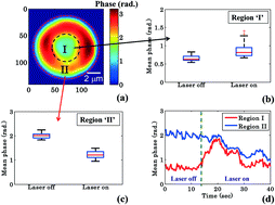
Red blood cells (RBCs) have the ability to undergo morphological deformations during microcirculation, such as changes in surface area, volume and sphericity. Optical waveguide trapping is suitable for trapping, propelling and deforming large cell populations along the length of the waveguide. Bright field microscopy employed with waveguide trapping does not provide quantitative information about structural changes. Here, we have combined quantitative phase microscopy and waveguide trapping techniques to study changes in RBC morphology during planar trapping and transportation. By using interference microscopy, time-lapsed interferometric images of trapped RBCs were recorded in real-time and subsequently utilized to reconstruct optical phase maps. Quantification of the phase differences before and after trapping enabled study of the mechanical effects during planar trapping. During planar trapping, a decrease in the maximum phase values, an increase in the surface area and a decrease in the volume and sphericity of RBCs were observed. QPM was used to analyze the phase values for two specific regions within RBCs: the annular rim and the central donut. The phase value of the annular rim decreases whereas it increases for the central donut during planar trapping. These changes correspond to a redistribution of cytosol inside the RBC during planar trapping and transportation.
中文翻译:

平面捕获和推进过程中红细胞的定量相显微镜观察†
红细胞 (RBC) 在微循环过程中能够发生形态变形,例如表面积、体积和球形度的变化。光波导捕获适用于沿波导长度捕获、推进和变形大细胞群。与波导捕获一起使用的明场显微镜不能提供有关结构变化的定量信息。在这里,我们结合了定量相显微镜和波导捕获技术来研究平面捕获和运输过程中红细胞形态的变化。通过使用干涉显微镜,实时记录捕获的红细胞的延时干涉图像,随后用于重建光学相位图。对捕获前后的相位差进行量化可以研究平面捕获期间的机械效应。在平面捕获过程中,观察到红细胞最大相位值下降、表面积增加以及体积和球形度下降。QPM 用于分析红细胞内两个特定区域的相位值:环形边缘和中央甜甜圈。在平面捕获期间,环形边缘的相位值减小,而中心圆环的相位值增大。这些变化对应于平面捕获和运输过程中红细胞内胞质的重新分布。
更新日期:2018-08-22
中文翻译:
平面捕获和推进过程中红细胞的定量相显微镜观察†
红细胞 (RBC) 在微循环过程中能够发生形态变形,例如表面积、体积和球形度的变化。光波导捕获适用于沿波导长度捕获、推进和变形大细胞群。与波导捕获一起使用的明场显微镜不能提供有关结构变化的定量信息。在这里,我们结合了定量相显微镜和波导捕获技术来研究平面捕获和运输过程中红细胞形态的变化。通过使用干涉显微镜,实时记录捕获的红细胞的延时干涉图像,随后用于重建光学相位图。对捕获前后的相位差进行量化可以研究平面捕获期间的机械效应。在平面捕获过程中,观察到红细胞最大相位值下降、表面积增加以及体积和球形度下降。QPM 用于分析红细胞内两个特定区域的相位值:环形边缘和中央甜甜圈。在平面捕获期间,环形边缘的相位值减小,而中心圆环的相位值增大。这些变化对应于平面捕获和运输过程中红细胞内胞质的重新分布。


 京公网安备 11010802027423号