Our official English website, www.x-mol.net, welcomes your feedback! (Note: you will need to create a separate account there.)
Nanoparticle localization in blood vessels: dependence on fluid shear stress, flow disturbances, and flow-induced changes in endothelial physiology†
Nanoscale ( IF 6.7 ) Pub Date : 2018-07-30 00:00:00 , DOI: 10.1039/c8nr03440k M. Juliana Gomez-Garcia 1, 2, 3 , Amber L. Doiron 2, 3, 4 , Robyn R. M. Steele 2, 3, 4 , Hagar I. Labouta 2, 5, 6, 7, 8 , Bahareh Vafadar 2, 3, 9 , Robert D. Shepherd 1, 2, 3 , Ian D. Gates 2, 3, 4 , David T. Cramb 2, 3, 8 , Sarah J. Childs 2, 3, 10 , Kristina D. Rinker 1, 2, 2, 3, 4
Nanoscale ( IF 6.7 ) Pub Date : 2018-07-30 00:00:00 , DOI: 10.1039/c8nr03440k M. Juliana Gomez-Garcia 1, 2, 3 , Amber L. Doiron 2, 3, 4 , Robyn R. M. Steele 2, 3, 4 , Hagar I. Labouta 2, 5, 6, 7, 8 , Bahareh Vafadar 2, 3, 9 , Robert D. Shepherd 1, 2, 3 , Ian D. Gates 2, 3, 4 , David T. Cramb 2, 3, 8 , Sarah J. Childs 2, 3, 10 , Kristina D. Rinker 1, 2, 2, 3, 4
Affiliation
|
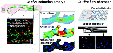
Nanoparticles in the bloodstream are subjected to complex fluid forces as they move through the curves and branches of healthy or tumor vasculature. While nanoparticles are known to preferentially accumulate in angiogenic vessels, little is known about the flow conditions in these vessels and how these conditions may influence localization. Here, we report a methodology which combines confocal imaging of nanoparticle-injected transgenic zebrafish embryos, 3D modeling of the vasculature, particle mapping, and computational fluid dynamics, to quantitatively assess the effects of fluid forces on nanoparticle distribution in vivo. Six-fold lower accumulation was found in zebrafish arteries compared to the lower velocity veins. Nanoparticle localization varied inversely with shear stress. Highest accumulation was present in regions of disturbed flow found at branch points and curvatures in the vasculature. To further investigate cell–particle association under flow, human endothelial cells were exposed to nanoparticles under hemodynamic conditions typically found in human vessels. Physiological adaptations of endothelial cells to 20 hours of flow enhanced nanoparticle accumulation in regions of disturbed flow. Overall our results suggest that fluid shear stress magnitude, flow disturbances, and flow-induced changes in endothelial physiology modulate nanoparticle localization in angiogenic vessels.
中文翻译:

纳米粒子在血管中的位置:依赖于流体剪切应力,流量干扰和流量诱导的内皮生理变化†
血液中的纳米粒子在穿过健康或肿瘤脉管系统的曲线和分支时会受到复杂的流体力。尽管已知纳米颗粒优先在血管生成血管中积聚,但对于这些血管中的血流状况以及这些状况如何影响定位知之甚少。在这里,我们报告了一种方法,该方法结合了注入纳米粒子的转基因斑马鱼胚胎的共聚焦成像,脉管系统的3D建模,粒子映射和计算流体动力学,以定量评估流体力对体内纳米粒子分布的影响。与低速静脉相比,斑马鱼动脉中的低六倍积累。纳米粒子的位置与剪切应力成反比。在脉管系统的分支点和弯曲处发现的紊流区域中存在最高的积累。为了进一步研究流动状态下的细胞-颗粒结合,人类血管内皮细胞在通常在人体血管中发现的血液动力学条件下暴露于纳米颗粒。内皮细胞对20小时血流的生理适应性增强了纳米颗粒在血流紊乱区域的积累。总的来说,我们的研究结果表明,流体剪切应力的大小,流动扰动和内皮生理中流动引起的变化可调节血管生成血管中的纳米颗粒定位。
更新日期:2018-07-30
中文翻译:
纳米粒子在血管中的位置:依赖于流体剪切应力,流量干扰和流量诱导的内皮生理变化†
血液中的纳米粒子在穿过健康或肿瘤脉管系统的曲线和分支时会受到复杂的流体力。尽管已知纳米颗粒优先在血管生成血管中积聚,但对于这些血管中的血流状况以及这些状况如何影响定位知之甚少。在这里,我们报告了一种方法,该方法结合了注入纳米粒子的转基因斑马鱼胚胎的共聚焦成像,脉管系统的3D建模,粒子映射和计算流体动力学,以定量评估流体力对体内纳米粒子分布的影响。与低速静脉相比,斑马鱼动脉中的低六倍积累。纳米粒子的位置与剪切应力成反比。在脉管系统的分支点和弯曲处发现的紊流区域中存在最高的积累。为了进一步研究流动状态下的细胞-颗粒结合,人类血管内皮细胞在通常在人体血管中发现的血液动力学条件下暴露于纳米颗粒。内皮细胞对20小时血流的生理适应性增强了纳米颗粒在血流紊乱区域的积累。总的来说,我们的研究结果表明,流体剪切应力的大小,流动扰动和内皮生理中流动引起的变化可调节血管生成血管中的纳米颗粒定位。


 京公网安备 11010802027423号