Our official English website, www.x-mol.net, welcomes your feedback! (Note: you will need to create a separate account there.)
Deep non-contact photoacoustic initial pressure imaging
Optica ( IF 10.4 ) Pub Date : 2018-07-09 , DOI: 10.1364/optica.5.000814 Parsin Haji Reza , Kevan Bell , Wei Shi , James Shapiro , Roger J. Zemp
Optica ( IF 10.4 ) Pub Date : 2018-07-09 , DOI: 10.1364/optica.5.000814 Parsin Haji Reza , Kevan Bell , Wei Shi , James Shapiro , Roger J. Zemp
|
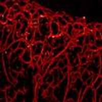
Photoacoustic imaging techniques have been extensively developed for biomedical applications, including functional and molecular imaging, due in part to their high optical contrast, high spatial resolution, and non-ionizing imaging properties. However, there are currently depth limitations in cellular-resolution, optically focused photoacoustic microscopy systems. In addition, most common photoacoustic systems need to be in contact with the sample through an ultrasound medium. In this work, by taking advantage of large photoacoustic initial pressures, all-optical non-contact optical resolution photoacoustic imaging is reported at depths beyond the optical transport mean-free path of the excitation wavelength. The proposed technique is called deep photoacoustic remote sensing (dPARS) microscopy. Visible pulsed excitation wavelengths are used to produce large initial-pressure-induced refractive index modulations in absorbing targets. These localized pressure rises create transient variations to the local scattering properties, which are detected as back-reflected intensity modulations from a deep-penetrating interrogation beam and do not require an interferometric detection pathway. Experiments demonstrate that dPARS is capable of providing optical resolution images to depths of 2.5 mm in tissue-mimicking scattering media. Signal-to-noise ratio is reported for in vivo imaging of microvascular networks. Also, imaging of single red blood cells, oxygen saturation mapping, and deep-vascular imaging applications are demonstrated. dPARS’s capabilities such as remote sensing, deep optical resolution imaging, and high signal-to-noise ratio, may yield new opportunities for several pre-clinical and clinical applications.
中文翻译:

深度非接触光声初始压力成像
光声成像技术已被广泛开发用于生物医学应用,包括功能和分子成像,这部分是由于它们的高光学对比度,高空间分辨率和非电离成像特性。然而,目前在细胞分辨率,光学聚焦的光声显微镜系统中存在深度限制。另外,大多数普通的光声系统需要通过超声介质与样品接触。在这项工作中,通过利用较大的光声初始压力,在超出激发波长的光传输无均值路径的深度报道了全光学非接触式光学分辨率光声成像。所提出的技术称为深光声遥感(dPARS)显微镜。可见的脉冲激发波长用于在吸收目标中产生大的初始压力诱导的折射率调制。这些局部压力上升会导致局部散射特性发生瞬态变化,这些变化会被深穿透探询光束作为背向反射强度调制进行检测,并且不需要干涉检测路径。实验表明,dPARS能够在模仿组织的散射介质中提供2.5毫米深度的光学分辨率图像。信噪比 它们被检测为来自深穿透询问光束的后向反射强度调制,并且不需要干涉检测路径。实验表明,dPARS能够在模仿组织的散射介质中提供2.5毫米深度的光学分辨率图像。信噪比 它们被检测为来自深穿透询问光束的后向反射强度调制,并且不需要干涉检测路径。实验表明,dPARS能够在模仿组织的散射介质中提供2.5毫米深度的光学分辨率图像。信噪比据报道用于微血管网络的体内成像。此外,还展示了单个红细胞的成像,氧饱和度作图和深层血管成像应用。dPARS的功能,例如遥感,深光学分辨率成像和高信噪比,可能会为一些临床前和临床应用带来新的机遇。
更新日期:2018-07-21
中文翻译:
深度非接触光声初始压力成像
光声成像技术已被广泛开发用于生物医学应用,包括功能和分子成像,这部分是由于它们的高光学对比度,高空间分辨率和非电离成像特性。然而,目前在细胞分辨率,光学聚焦的光声显微镜系统中存在深度限制。另外,大多数普通的光声系统需要通过超声介质与样品接触。在这项工作中,通过利用较大的光声初始压力,在超出激发波长的光传输无均值路径的深度报道了全光学非接触式光学分辨率光声成像。所提出的技术称为深光声遥感(dPARS)显微镜。可见的脉冲激发波长用于在吸收目标中产生大的初始压力诱导的折射率调制。这些局部压力上升会导致局部散射特性发生瞬态变化,这些变化会被深穿透探询光束作为背向反射强度调制进行检测,并且不需要干涉检测路径。实验表明,dPARS能够在模仿组织的散射介质中提供2.5毫米深度的光学分辨率图像。信噪比 它们被检测为来自深穿透询问光束的后向反射强度调制,并且不需要干涉检测路径。实验表明,dPARS能够在模仿组织的散射介质中提供2.5毫米深度的光学分辨率图像。信噪比 它们被检测为来自深穿透询问光束的后向反射强度调制,并且不需要干涉检测路径。实验表明,dPARS能够在模仿组织的散射介质中提供2.5毫米深度的光学分辨率图像。信噪比据报道用于微血管网络的体内成像。此外,还展示了单个红细胞的成像,氧饱和度作图和深层血管成像应用。dPARS的功能,例如遥感,深光学分辨率成像和高信噪比,可能会为一些临床前和临床应用带来新的机遇。


 京公网安备 11010802027423号