Our official English website, www.x-mol.net, welcomes your feedback! (Note: you will need to create a separate account there.)
3D cell electrorotation and imaging for measuring multiple cellular biophysical properties†
Lab on a Chip ( IF 6.1 ) Pub Date : 2018-06-18 00:00:00 , DOI: 10.1039/c8lc00407b Liang Huang 1, 2, 3, 4, 5 , Peng Zhao 1, 2, 3, 4, 5 , Wenhui Wang 1, 2, 3, 4, 5
Lab on a Chip ( IF 6.1 ) Pub Date : 2018-06-18 00:00:00 , DOI: 10.1039/c8lc00407b Liang Huang 1, 2, 3, 4, 5 , Peng Zhao 1, 2, 3, 4, 5 , Wenhui Wang 1, 2, 3, 4, 5
Affiliation

|
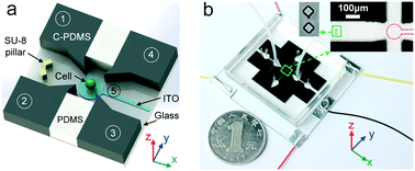
3D rotation is one of many fundamental manipulations to cells and imperative in a wide range of applications in single cell analysis involving biology, chemistry, physics and medicine. In this article, we report a dielectrophoresis-based, on-chip manipulation method that can load and rotate a single cell for 3D cell imaging and multiple biophysical property measurements. To achieve this, we trapped a single cell in constriction and subsequently released it to a rotation chamber formed by four sidewall electrodes and one transparent bottom electrode. In the rotation chamber, rotating electric fields were generated by applying appropriate AC signals to the electrodes for driving the single cell to rotate in 3D under control. The rotation spectrum for in-plane rotation was used to extract the cellular dielectric properties based on a spherical single-shell model, and the stacked images of out-of-plane cell rotation were used to reconstruct the 3D cell morphology to determine its geometric parameters. We have tested the capabilities of our method by rotating four representative mammalian cells including HeLa, C3H10, B lymphocyte, and HepaRG. Using our device, we quantified the area-specific membrane capacitance and cytoplasm conductivity for the four cells, and revealed the subtle difference of geometric parameters (i.e., surface area, volume, and roughness) by 3D cell imaging of cancer cells and normal leukocytes. Combining microfluidics, dielectrophoresis, and microscopic imaging techniques, our electrorotation-on-chip (EOC) technique is a versatile method for manipulating single cells under investigation and measuring their multiple biophysical properties.
中文翻译:

用于测量多种细胞生物物理特性的3D细胞电旋转和成像†
3D旋转是对细胞进行的许多基本操作之一,在涉及生物学,化学,物理学和医学的单细胞分析的广泛应用中,当务之急是3D旋转。在本文中,我们报告了一种基于介电电泳的片上操作方法,该方法可以加载和旋转单个细胞以进行3D细胞成像和多个生物物理性质测量。为了实现这一目标,我们将单个电池陷在狭窄处,然后将其释放到由四个侧壁电极和一个透明底部电极形成的旋转腔中。在旋转室中,通过向电极施加适当的AC信号来产生旋转电场,以驱动单个单元在控制下以3D旋转。平面内旋转的旋转光谱用于基于球形单壳模型提取细胞介电特性,平面外单元旋转的堆叠图像用于重建3D单元形态,以确定其几何参数。我们已经通过旋转四个代表性的哺乳动物细胞(包括HeLa,C3H10,B淋巴细胞和HepaRG)来测试了我们方法的功能。使用我们的设备,我们量化了这四个细胞的区域特异性膜电容和细胞质电导率,并揭示了几何参数之间的细微差异(我们已经通过旋转四个代表性的哺乳动物细胞(包括HeLa,C3H10,B淋巴细胞和HepaRG)来测试了我们方法的功能。使用我们的设备,我们量化了这四个细胞的区域特异性膜电容和细胞质电导率,并揭示了几何参数之间的细微差异(我们已经通过旋转四个代表性的哺乳动物细胞(包括HeLa,C3H10,B淋巴细胞和HepaRG)来测试了我们方法的功能。使用我们的设备,我们量化了这四个细胞的区域特异性膜电容和细胞质电导率,并揭示了几何参数之间的细微差异((例如,表面积,体积和粗糙度)通过癌细胞和正常白细胞的3D细胞成像进行。结合微流体技术,介电电泳技术和显微成像技术,我们的芯片电旋转(EOC)技术是一种用于处理单个细胞并测量其多种生物物理特性的通用方法。
更新日期:2018-06-18
中文翻译:

用于测量多种细胞生物物理特性的3D细胞电旋转和成像†
3D旋转是对细胞进行的许多基本操作之一,在涉及生物学,化学,物理学和医学的单细胞分析的广泛应用中,当务之急是3D旋转。在本文中,我们报告了一种基于介电电泳的片上操作方法,该方法可以加载和旋转单个细胞以进行3D细胞成像和多个生物物理性质测量。为了实现这一目标,我们将单个电池陷在狭窄处,然后将其释放到由四个侧壁电极和一个透明底部电极形成的旋转腔中。在旋转室中,通过向电极施加适当的AC信号来产生旋转电场,以驱动单个单元在控制下以3D旋转。平面内旋转的旋转光谱用于基于球形单壳模型提取细胞介电特性,平面外单元旋转的堆叠图像用于重建3D单元形态,以确定其几何参数。我们已经通过旋转四个代表性的哺乳动物细胞(包括HeLa,C3H10,B淋巴细胞和HepaRG)来测试了我们方法的功能。使用我们的设备,我们量化了这四个细胞的区域特异性膜电容和细胞质电导率,并揭示了几何参数之间的细微差异(我们已经通过旋转四个代表性的哺乳动物细胞(包括HeLa,C3H10,B淋巴细胞和HepaRG)来测试了我们方法的功能。使用我们的设备,我们量化了这四个细胞的区域特异性膜电容和细胞质电导率,并揭示了几何参数之间的细微差异(我们已经通过旋转四个代表性的哺乳动物细胞(包括HeLa,C3H10,B淋巴细胞和HepaRG)来测试了我们方法的功能。使用我们的设备,我们量化了这四个细胞的区域特异性膜电容和细胞质电导率,并揭示了几何参数之间的细微差异((例如,表面积,体积和粗糙度)通过癌细胞和正常白细胞的3D细胞成像进行。结合微流体技术,介电电泳技术和显微成像技术,我们的芯片电旋转(EOC)技术是一种用于处理单个细胞并测量其多种生物物理特性的通用方法。


 京公网安备 11010802027423号