Our official English website, www.x-mol.net, welcomes your feedback! (Note: you will need to create a separate account there.)
Scan-less confocal phase imaging based on dual-comb microscopy
Optica ( IF 10.4 ) Pub Date : 2018-05-16 , DOI: 10.1364/optica.5.000634 Eiji Hase , Takeo Minamikawa , Takahiko Mizuno , Shuji Miyamoto , Ryuji Ichikawa , Yi-Da Hsieh , Kyuki Shibuya , Katsuya Sato , Yoshiaki Nakajima , Akifumi Asahara , Kaoru Minoshima , Yasuhiro Mizutani , Tetsuo Iwata , Hirotsugu Yamamoto , Takeshi Yasui
Optica ( IF 10.4 ) Pub Date : 2018-05-16 , DOI: 10.1364/optica.5.000634 Eiji Hase , Takeo Minamikawa , Takahiko Mizuno , Shuji Miyamoto , Ryuji Ichikawa , Yi-Da Hsieh , Kyuki Shibuya , Katsuya Sato , Yoshiaki Nakajima , Akifumi Asahara , Kaoru Minoshima , Yasuhiro Mizutani , Tetsuo Iwata , Hirotsugu Yamamoto , Takeshi Yasui
|
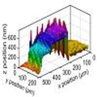
Confocal laser microscopy (CLM) is a powerful tool in life science research and industrial inspection because it offers two-dimensional optical sectioning or three-dimensional imaging capability with micrometer depth selectivity. Furthermore, scan-less imaging modality enables rapid image acquisition and high robustness against surrounding external disturbances in CLM. However, the objects to be measured must be reflective, absorptive, scattering, or fluorescent because the image contrast is given by the optical intensity. If a new image contrast can be provided by the optical phase, scan-less CLM can be further applied for transparent non-fluorescent objects or reflective objects with nanometer unevenness by providing information on refractive index, optical thickness, or geometrical shape. Here, we report scan-less confocal dual-comb microscopy offering a phase image in addition to an amplitude image with depth selectivity by using an optical frequency comb as an optical carrier of amplitude and phase with discrete ultra-multichannels. Our technique encodes confocal amplitude and phase images of a sample onto a series of discrete modes in the optical frequency comb with well-defined amplitude and phase to establish a one-to-one correspondence between image pixels and comb modes. The technique then decodes these images from comb modes with amplitude and phase. We demonstrate confocal phase imaging with milliradian phase resolution under micrometer depth selectivity on the millisecond timescale. As a proof of concept, we demonstrate the quantitative phase imaging of standing culture fixed cells and the surface topography of nanometer-scale step structures. Our technique for confocal phase imaging will find applications in three-dimensional visualization of stacked living cells in culture and nanometer surface topography of semiconductor objects.
中文翻译:

基于双梳显微镜的无扫描共聚焦相成像
共聚焦激光显微镜(CLM)是生命科学研究和工业检查中的强大工具,因为它提供了具有微米深度选择性的二维光学切片或三维成像功能。此外,无扫描成像方式可实现快速图像采集和针对CLM中周围周围外部干扰的高鲁棒性。但是,要测量的物体必须是反射性,吸收性,散射性或荧光性的,因为图像对比度是由光强度给出的。如果可以通过光相提供新的图像对比度,则可以通过提供有关折射率,光学厚度或几何形状的信息,将无扫描CLM进一步应用于具有纳米凹凸的透明非荧光对象或反射对象。这里,我们报道了通过使用光学频率梳作为具有离散超多通道的振幅和相位的光学载体,无扫描共聚焦双梳形显微镜除了提供具有深度选择性的振幅图像外,还提供相位图像。我们的技术将样本的共焦振幅和相位图像编码到光学频率梳中具有明确定义的振幅和相位的一系列离散模式,以在图像像素和梳状模式之间建立一对一的对应关系。然后,该技术从具有幅度和相位的梳状模式中解码这些图像。我们演示了在毫秒时间尺度上在微米深度选择性下具有弧度相位分辨率的共焦相成像。作为概念证明,我们展示了静置培养固定细胞的定量相成像和纳米级台阶结构的表面形貌。我们的共聚焦相成像技术将在堆叠活细胞的三维可视化以及半导体物体的纳米表面形貌的三维可视化中找到应用。
更新日期:2018-05-18
中文翻译:
基于双梳显微镜的无扫描共聚焦相成像
共聚焦激光显微镜(CLM)是生命科学研究和工业检查中的强大工具,因为它提供了具有微米深度选择性的二维光学切片或三维成像功能。此外,无扫描成像方式可实现快速图像采集和针对CLM中周围周围外部干扰的高鲁棒性。但是,要测量的物体必须是反射性,吸收性,散射性或荧光性的,因为图像对比度是由光强度给出的。如果可以通过光相提供新的图像对比度,则可以通过提供有关折射率,光学厚度或几何形状的信息,将无扫描CLM进一步应用于具有纳米凹凸的透明非荧光对象或反射对象。这里,我们报道了通过使用光学频率梳作为具有离散超多通道的振幅和相位的光学载体,无扫描共聚焦双梳形显微镜除了提供具有深度选择性的振幅图像外,还提供相位图像。我们的技术将样本的共焦振幅和相位图像编码到光学频率梳中具有明确定义的振幅和相位的一系列离散模式,以在图像像素和梳状模式之间建立一对一的对应关系。然后,该技术从具有幅度和相位的梳状模式中解码这些图像。我们演示了在毫秒时间尺度上在微米深度选择性下具有弧度相位分辨率的共焦相成像。作为概念证明,我们展示了静置培养固定细胞的定量相成像和纳米级台阶结构的表面形貌。我们的共聚焦相成像技术将在堆叠活细胞的三维可视化以及半导体物体的纳米表面形貌的三维可视化中找到应用。


 京公网安备 11010802027423号