Analytical and Bioanalytical Chemistry ( IF 4.3 ) Pub Date : 2018-03-17 , DOI: 10.1007/s00216-018-0987-9 Katia Wehbe , Marzia Vezzalini , Gianfelice Cinque
Mycoplasma contamination represents a significant problem to the culture of mammalian cells used for research as it can cause disastrous effects on eukaryotic cells by altering cellular parameters leading to unreliable experimental results. Mycoplasma cells are very small bacteria therefore they cannot be detected by visual inspection using a visible light microscope and, thus, can remain unnoticed in the cell cultures for long periods. The detection techniques used nowadays to reveal mycoplasma contamination are time consuming and expensive with each having significant drawbacks. The ideal detection should be simple to perform with minimal preparation time, rapid, inexpensive, and sensitive. To our knowledge, for the first time, we employed Fourier transform infrared (FTIR) microspectroscopy to investigate whether we can differentiate between control cells and the same cells which have been infected with mycoplasmas during the culturing process. Chemometric methods such as HCA and PCA were used for the data analysis in order to detect spectral differences between control and intentionally infected cells, and spectral markers were revealed even at low contamination level. The preliminary results showed that FTIR has the potential to be used in the future as a reliable complementary detection technique for mycoplasma-infected cells.
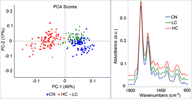
FTIR microspectroscopy is able to differentiate between mycoplasma infected cells (LC for low contamination and HC for high contamination) and control non-infected cells (CN).
中文翻译:
使用FTIR显微技术检测受污染的哺乳动物细胞培养物中的支原体
支原体污染对于用于研究的哺乳动物细胞培养是一个重大问题,因为它会通过改变细胞参数导致不可靠的实验结果而对真核细胞造成灾难性影响。支原体细胞是非常小的细菌,因此无法通过使用可见光显微镜的目测进行检测,因此很长一段时间都不会在细胞培养物中引起注意。当今用于揭示支原体污染的检测技术既耗时又昂贵,每种技术都有明显的缺陷。理想的检测应易于执行,准备时间最少,快速,廉价且灵敏。据我们所知,这是第一次 我们使用傅里叶变换红外(FTIR)显微技术研究了我们能否在培养过程中区分对照细胞和感染支原体的相同细胞。为了检测对照细胞和故意感染细胞之间的光谱差异,使用了化学计量学方法(例如HCA和PCA)进行数据分析,即使在低污染水平下也能显示光谱标记。初步结果表明,FTIR有可能在将来用作支原体感染细胞的可靠补充检测技术。为了检测对照细胞和故意感染细胞之间的光谱差异,使用了化学计量学方法(例如HCA和PCA)进行数据分析,即使在低污染水平下也能显示光谱标记。初步结果表明,FTIR有可能在将来用作支原体感染细胞的可靠补充检测技术。为了检测对照细胞和故意感染细胞之间的光谱差异,使用了化学计量学方法(例如HCA和PCA)进行数据分析,即使在低污染水平下也能显示光谱标记。初步结果表明,FTIR有可能在将来用作支原体感染细胞的可靠补充检测技术。
FTIR显微技术能够区分支原体感染的细胞(低污染的是LC,高污染的是HC)并控制了未感染的细胞(CN)。


 京公网安备 11010802027423号