当前位置:
X-MOL 学术
›
Adv. Funct. Mater.
›
论文详情
Our official English website, www.x-mol.net, welcomes your feedback! (Note: you will need to create a separate account there.)
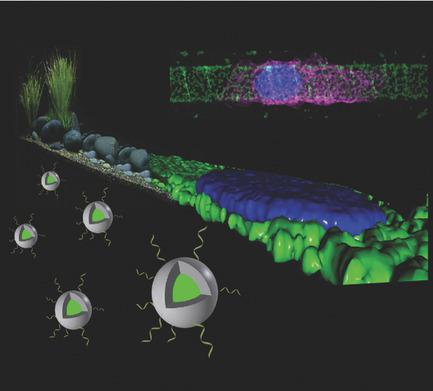
Biopebbles: DNA‐Functionalized Core–Shell Silica Nanospheres for Cellular Uptake and Cell Guidance Studies
Advanced Functional Materials ( IF 19.0 ) Pub Date : 2018-03-09 , DOI: 10.1002/adfm.201707572 Arnold Leidner 1 , Simone Weigel 1 , Jens Bauer 1 , Jens Reiber 1, 2 , Alessandro Angelin 1 , Maximilian Grösche 1 , Tim Scharnweber 1 , Christof M. Niemeyer 1
Advanced Functional Materials ( IF 19.0 ) Pub Date : 2018-03-09 , DOI: 10.1002/adfm.201707572 Arnold Leidner 1 , Simone Weigel 1 , Jens Bauer 1 , Jens Reiber 1, 2 , Alessandro Angelin 1 , Maximilian Grösche 1 , Tim Scharnweber 1 , Christof M. Niemeyer 1
Affiliation
|
The development of a versatile class of silica nanoparticles for cell studies is reported. The particles contain a fluorescent dye‐encoded core and a single‐stranded DNA oligonucleotide‐displaying shell. They are accessible in arbitrary size and color through robust protocols for Stöber‐based colloidal synthesis and sturdy chemical surface functionalization. Silica particles in the size range of 100 nm to 1.5 µm diameter containing fluorescein, Cy3 oder Cy5 dye‐encoded cores are synthesized and functionalized with DNA oligonucleotides. These silica biopebbles are conveniently traceable by microscopy and have a high affinity to live cells, which makes them ideal for cell uptake studies, as demonstrated for MCF7 and A431 cancer cells. The biopebbles can be utilized as building blocks for the self‐assembled formation of arbitrary surface patterns on glass substrates. With these architectures, the privileged internalization of the biopebbles can be exploited for improved adhesion and guidance of cells because the particles are no longer ingested by adhered cells due to their physical connection with the solid support. It is believed that the biopebble approach will be useful for a variety of applications, fundamental studies in cell biology and tissue engineering.
中文翻译:

生物卵石:DNA功能化的核壳二氧化硅纳米球用于细胞吸收和细胞指导研究
据报道,用于细胞研究的一类通用的二氧化硅纳米颗粒的开发。颗粒包含荧光染料编码的核心和单链DNA寡核苷酸展示壳。通过基于Stöber的胶体合成和坚固的化学表面功能化的强大协议,可以以任意大小和颜色访问它们。合成了直径范围为100 nm至1.5 µm的含荧光素的二氧化硅颗粒,并用DNA寡核苷酸对Cy3或Cy5染料编码的核进行了功能化。这些二氧化硅生物卵石可通过显微镜方便地追踪到,并且对活细胞具有高亲和力,这使其成为细胞摄取研究的理想选择,如MCF7和A431癌细胞所证明的。生物卵石可以用作在玻璃基板上自组装形成任意表面图案的基础。利用这些结构,可以利用生物卵石的特权内部化来改善细胞的粘附和引导,因为由于其与固体支持物的物理连接,颗粒不再被粘附的细胞摄取。相信生物卵石方法将对细胞生物学和组织工程学的各种应用,基础研究有用。
更新日期:2018-03-09
中文翻译:
生物卵石:DNA功能化的核壳二氧化硅纳米球用于细胞吸收和细胞指导研究
据报道,用于细胞研究的一类通用的二氧化硅纳米颗粒的开发。颗粒包含荧光染料编码的核心和单链DNA寡核苷酸展示壳。通过基于Stöber的胶体合成和坚固的化学表面功能化的强大协议,可以以任意大小和颜色访问它们。合成了直径范围为100 nm至1.5 µm的含荧光素的二氧化硅颗粒,并用DNA寡核苷酸对Cy3或Cy5染料编码的核进行了功能化。这些二氧化硅生物卵石可通过显微镜方便地追踪到,并且对活细胞具有高亲和力,这使其成为细胞摄取研究的理想选择,如MCF7和A431癌细胞所证明的。生物卵石可以用作在玻璃基板上自组装形成任意表面图案的基础。利用这些结构,可以利用生物卵石的特权内部化来改善细胞的粘附和引导,因为由于其与固体支持物的物理连接,颗粒不再被粘附的细胞摄取。相信生物卵石方法将对细胞生物学和组织工程学的各种应用,基础研究有用。


 京公网安备 11010802027423号