当前位置:
X-MOL 学术
›
Environ. Sci. Technol. Lett.
›
论文详情
Our official English website, www.x-mol.net, welcomes your feedback! (Note: you will need to create a separate account there.)
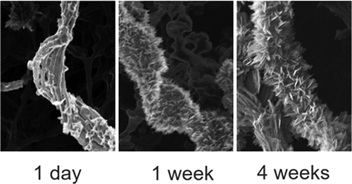
Imaging Organic–Mineral Aggregates Formed by Fe(II)-Oxidizing Bacteria Using Helium Ion Microscopy
Environmental Science & Technology Letters ( IF 10.9 ) Pub Date : 2018-03-07 00:00:00 , DOI: 10.1021/acs.estlett.8b00077 James M. Byrne 1 , Matthias Schmidt 2 , Tina Gauger 1 , Casey Bryce 1 , Andreas Kappler 1
Environmental Science & Technology Letters ( IF 10.9 ) Pub Date : 2018-03-07 00:00:00 , DOI: 10.1021/acs.estlett.8b00077 James M. Byrne 1 , Matthias Schmidt 2 , Tina Gauger 1 , Casey Bryce 1 , Andreas Kappler 1
Affiliation
|
Helium ion microscopy (HIM) has been used to image the development of mineralized twisted stalks produced by a neutrophilic, microaerophilic Fe(II)-oxidizing bacteria of the class Zetaproteobacteria. HIM is a relatively new type of microscopy which has several advantages over conventional scanning electron microscopy (SEM) due to its higher spatial resolution and the fact that samples can be imaged without coating (e.g., with Pt/Au). Here, we use HIM to show the development of nanometer- and micrometer-sized twisted stalk features consisting of organic material and Fe(III) minerals which appear to be loosely bound to the bacterial cells. These appendages are thought to be essential for eliminating Fe(III) waste produced during Fe(II) oxidation by sorbing Fe(III) and transporting it away from the cell. The results show the initial formation of long, precipitate-free stalks. After just 2 days, these became encrusted with spiky lepidocrocite crystals, and by 1 month, the characteristic twisted shape was almost indiscernible. These results demonstrate the high quality of images which can be obtained with helium ion microscopy from organic and mineral structures produced by bacteria without the requirement to sputter coat samples with conductive metals and can thus be considered to be more representive of how these structures would exist in the environment.
中文翻译:

使用氦离子显微镜对Fe(II)氧化细菌形成的有机矿物质聚集体进行成像
氦离子显微镜(HIM)已用于成像由Zetaproteobacteria类嗜中性,微需氧性Fe(II)氧化细菌产生的矿化扭曲茎的发育过程。HIM是一种相对较新的显微镜,由于其较高的空间分辨率和无需涂层即可成像(例如,使用Pt / Au成像)的事实,与常规的扫描电子显微镜(SEM)相比,它具有多个优势。在这里,我们使用HIM展示了由有机材料和Fe(III)矿物质组成的纳米级和微米级扭曲茎杆特征的发展,这些矿物似乎松散地结合在细菌细胞上。这些附属物被认为对于通过吸附Fe(III)并将其运离电池而消除Fe(II)氧化过程中产生的Fe(III)废物至关重要。结果表明,最初形成了长而无沉淀的茎。仅用了2天,这些材料就被尖尖的铁云母晶体包裹住了,到1个月时,特征扭曲的形状几乎变得难以辨认。
更新日期:2018-03-08
中文翻译:
使用氦离子显微镜对Fe(II)氧化细菌形成的有机矿物质聚集体进行成像
氦离子显微镜(HIM)已用于成像由Zetaproteobacteria类嗜中性,微需氧性Fe(II)氧化细菌产生的矿化扭曲茎的发育过程。HIM是一种相对较新的显微镜,由于其较高的空间分辨率和无需涂层即可成像(例如,使用Pt / Au成像)的事实,与常规的扫描电子显微镜(SEM)相比,它具有多个优势。在这里,我们使用HIM展示了由有机材料和Fe(III)矿物质组成的纳米级和微米级扭曲茎杆特征的发展,这些矿物似乎松散地结合在细菌细胞上。这些附属物被认为对于通过吸附Fe(III)并将其运离电池而消除Fe(II)氧化过程中产生的Fe(III)废物至关重要。结果表明,最初形成了长而无沉淀的茎。仅用了2天,这些材料就被尖尖的铁云母晶体包裹住了,到1个月时,特征扭曲的形状几乎变得难以辨认。


 京公网安备 11010802027423号