当前位置:
X-MOL 学术
›
Mol. Pharmaceutics
›
论文详情
Our official English website, www.x-mol.net, welcomes your feedback! (Note: you will need to create a separate account there.)
Immuno-PET Imaging of 89Zr Labeled Anti-PD-L1 Domain Antibody
Molecular Pharmaceutics ( IF 4.9 ) Pub Date : 2018-03-05 00:00:00 , DOI: 10.1021/acs.molpharmaceut.8b00062 Dan Li 1 , Siyuan Cheng 1 , Sijuan Zou 1 , Dongling Zhu 1 , Tinghui Zhu 2 , Pilin Wang 2 , Xiaohua Zhu 1
Molecular Pharmaceutics ( IF 4.9 ) Pub Date : 2018-03-05 00:00:00 , DOI: 10.1021/acs.molpharmaceut.8b00062 Dan Li 1 , Siyuan Cheng 1 , Sijuan Zou 1 , Dongling Zhu 1 , Tinghui Zhu 2 , Pilin Wang 2 , Xiaohua Zhu 1
Affiliation
|
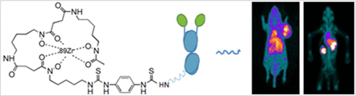
Recently, various immuno-PET tracers based on monoclonal antibodies (mAbs), engineered scaffold proteins, and peptides were developed to target either programmed cell death protein 1 (PD-1) or programmed cell death ligand 1 (PD-L1), showing promise in assessment of immune checkpoints. We sought to develop an immunotherapeutic agent based PET probe that enables real-time assessment of PD-L1 expression and evaluation of antibody drug biodistribution to select eligible candidates for anti-PD-1/PD-L1 immunotherapies. KN035, a 79.6 kDa size anti-PD-L1 domain antibody under analysis in clinical trials, was used to develop the immuno-PET probe, 89Zr-Df-KN035. Immuno-PET studies were performed to monitor PD-L1 levels in nude mice bearing LN229 xenografts with positive expression for PD-L1, and to evaluate the whole-body biodistribution in healthy non-human primates (NHPs). LN229 xenografts were markedly visualized from 24 h after injection of 89Zr-Df-KN035, with elevated accumulation persisting for up to 120 h. Tumor radioactivity was notably reduced in the presence of excess KN035. Mouse ex vivo biodistribution studies performed at 24 and 120 h revealed tumor-to-muscle ratios as high as 5.64 ± 0.65 and 7.70 ± 1.37, respectively. In the NHP model, PET imaging demonstrated low background. The liver and kidney showed moderate accumulation with the highest SUVmean value of 1.15 ± 0.15 and 2.13 ± 0.10 at 72 h, respectively. The spleen, lymph nodes, and salivary glands were also slightly visualized. In conclusion, 89Zr-Df-KN035, a novel anti-PD-L1 domain antibody-based probe, shows the feasibility of noninvasive in vivo evaluation of PD-L1 expression. This work further provides a template for immunotherapeutic agent based imaging to evaluate human PD-L1 expression and to augment our understanding of therapeutic agent biodistribution, leading to better therapeutic strategies in the future.
中文翻译:

89 Zr标记的抗PD-L1域抗体的免疫PET成像
最近,开发了多种基于单克隆抗体(mAbs),工程支架蛋白和肽的免疫PET示踪剂,以靶向程序性细胞死亡蛋白1(PD-1)或程序性细胞死亡配体1(PD-L1),显示出希望在评估免疫检查点。我们寻求开发一种基于免疫治疗剂的PET探针,该探针能够实时评估PD-L1的表达并评估抗体药物的生物分布,从而为抗PD-1 / PD-L1免疫疗法选择合适的候选药物。KN035是一种79.6 kDa大小的抗PD-L1结构域抗体,正在临床试验中进行分析,用于开发免疫PET探针89Zr-Df-KN035。进行了Immuno-PET研究,以监测荷有PD-L1阳性表达的LN229异种移植裸鼠的PD-L1水平,并评估健康人非灵长类动物(NHP)中的全身生物分布。LN229异种移植物从注射89 Zr-Df-KN035后的24 h显着可见,累积的积累持续长达120 h。在过量KN035的存在下,肿瘤放射性显着降低。在24和120 h进行的小鼠离体生物分布研究显示,肿瘤与肌肉的比例分别高达5.64±0.65和7.70±1.37。在NHP模型中,PET成像显示低背景。肝和肾显示中等程度的蓄积,SUV平均值最高72小时时的数值分别为1.15±0.15和2.13±0.10。脾脏,淋巴结和唾液腺也略显。总之,一种基于抗PD-L1结构域抗体的新型探针89 Zr-Df-KN035显示了非侵入性体内评估PD-L1表达的可行性。这项工作进一步为基于免疫治疗剂的成像提供了模板,以评估人PD-L1的表达并增强我们对治疗剂生物分布的理解,从而在将来带来更好的治疗策略。
更新日期:2018-03-05
中文翻译:
89 Zr标记的抗PD-L1域抗体的免疫PET成像
最近,开发了多种基于单克隆抗体(mAbs),工程支架蛋白和肽的免疫PET示踪剂,以靶向程序性细胞死亡蛋白1(PD-1)或程序性细胞死亡配体1(PD-L1),显示出希望在评估免疫检查点。我们寻求开发一种基于免疫治疗剂的PET探针,该探针能够实时评估PD-L1的表达并评估抗体药物的生物分布,从而为抗PD-1 / PD-L1免疫疗法选择合适的候选药物。KN035是一种79.6 kDa大小的抗PD-L1结构域抗体,正在临床试验中进行分析,用于开发免疫PET探针89Zr-Df-KN035。进行了Immuno-PET研究,以监测荷有PD-L1阳性表达的LN229异种移植裸鼠的PD-L1水平,并评估健康人非灵长类动物(NHP)中的全身生物分布。LN229异种移植物从注射89 Zr-Df-KN035后的24 h显着可见,累积的积累持续长达120 h。在过量KN035的存在下,肿瘤放射性显着降低。在24和120 h进行的小鼠离体生物分布研究显示,肿瘤与肌肉的比例分别高达5.64±0.65和7.70±1.37。在NHP模型中,PET成像显示低背景。肝和肾显示中等程度的蓄积,SUV平均值最高72小时时的数值分别为1.15±0.15和2.13±0.10。脾脏,淋巴结和唾液腺也略显。总之,一种基于抗PD-L1结构域抗体的新型探针89 Zr-Df-KN035显示了非侵入性体内评估PD-L1表达的可行性。这项工作进一步为基于免疫治疗剂的成像提供了模板,以评估人PD-L1的表达并增强我们对治疗剂生物分布的理解,从而在将来带来更好的治疗策略。


 京公网安备 11010802027423号