Acta Biomaterialia ( IF 9.7 ) Pub Date : 2018-03-02 , DOI: 10.1016/j.actbio.2018.02.030 J. Patel , S. Lal , K. Nuss , S.P. Wilshaw , B. von Rechenberg , R.M. Hall , J.L. Tipper
|
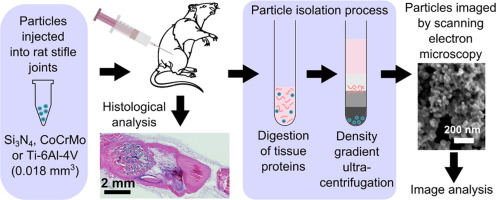
Less than optimal particle isolation techniques have impeded analysis of orthopaedic wear debris in vivo. The purpose of this research was to develop and test an improved method for particle isolation from tissue. A volume of 0.018 mm3 of clinically relevant CoCrMo, Ti-6Al-4V or Si3N4 particles was injected into rat stifle joints for seven days of in vivo exposure. Following sacrifice, particles were located within tissues using histology. The particles were recovered by enzymatic digestion of periarticular tissue with papain and proteinase K, followed by ultracentrifugation using a sodium polytungstate density gradient. Particles were recovered from all samples, observed using SEM and the particle composition was verified using EDX, which demonstrated that all isolated particles were free from contamination. Particle size, aspect ratio and circularity were measured using image analysis software. There were no significant changes to the measured parameters of CoCrMo or Si3N4 particles before and after the recovery process (KS tests, p>0.05). Titanium particles were too few before and after isolation to analyse statistically, though size and morphologies were similar. Overall the method demonstrated a significant improvement to current particle isolation methods from tissue in terms of sensitivity and efficacy at removal of protein, and has the potential to be used for the isolation of ultra-low wearing total joint replacement materials from periprosthetic tissues.
Statement of Significance
This research presents a novel method for the isolation of wear particles from tissue. Methodology outlined in this work would be a valuable resource for future researchers wishing to isolate particles from tissues, either as part of preclinical testing, or from explants from patients for diagnostic purposes. It is increasingly recognised that analysis of wear particles is critical to evaluating the safety of an orthopaedic device.
中文翻译:
使用新型颗粒分离方法从大鼠窒息关节组织中回收少量磨损碎片
不够理想的颗粒分离技术阻碍了骨科磨损碎片在体内的分析。这项研究的目的是开发和测试一种改进的从组织中分离颗粒的方法。在体内进行7天的注射后,将0.018 mm 3的临床相关CoCrMo,Ti-6Al-4V或Si 3 N 4颗粒注入大鼠的窒息关节中接触。处死后,使用组织学将颗粒定位在组织内。通过用木瓜蛋白酶和蛋白酶K对关节周围组织进行酶消化,然后使用聚钨酸钠密度梯度进行超速离心来回收颗粒。从所有样品中回收颗粒,使用SEM观察,并使用EDX验证颗粒组成,这表明所有分离出的颗粒均无污染。使用图像分析软件测量粒度,长宽比和圆度。CoCrMo或Si 3 N 4的测量参数没有明显变化恢复过程前后的颗粒(KS试验,p> 0.05)。钛粒子分离前后太少,无法进行统计分析,尽管尺寸和形貌相似。总体而言,该方法在去除蛋白质方面的敏感性和功效方面,证明了从组织中分离当前粒子的方法有了重大改进,并且有潜力用于从假体周围组织中分离超低磨损的全关节置换材料。
重要声明
这项研究提出了一种从组织中分离磨损颗粒的新方法。对于将来希望从组织中分离出颗粒(作为临床前测试的一部分)或从患者的外植体中分离出来以进行诊断的研究人员而言,这项工作中概述的方法学将是宝贵的资源。人们越来越认识到,磨损颗粒的分析对于评估骨科器械的安全性至关重要。


 京公网安备 11010802027423号