Our official English website, www.x-mol.net, welcomes your feedback! (Note: you will need to create a separate account there.)
Spatio-Temporal Image Correlation Spectroscopy and Super-Resolution Microscopy to Quantify Molecular Dynamics in T cells
Methods ( IF 4.8 ) Pub Date : 2018-05-01 , DOI: 10.1016/j.ymeth.2018.01.017 George W. Ashdown , Dylan M. Owen
Methods ( IF 4.8 ) Pub Date : 2018-05-01 , DOI: 10.1016/j.ymeth.2018.01.017 George W. Ashdown , Dylan M. Owen
|
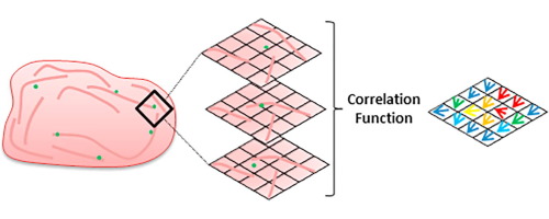
Many cellular processes are regulated by the spatio-temporal organisation of signalling complexes, cytoskeletal components and membranes. One such example is at the T cell immunological synapse where the retrograde flow of cortical filamentous (F)-actin from the synapse periphery drives signalling protein microclusters towards the synapse centre. The density of this mesh however, makes visualisation and analysis of individual actin fibres difficult due to the resolution limit of conventional microscopy. Recently, super-resolution methods such as structured illumination microscopy (SIM) have surpassed this resolution limit. Here, we apply SIM to better visualise the dense cortical actin meshwork in T cell synapses formed against activating, antibody-coated surfaces and image under total-internal reflection fluorescence (TIRF) illumination. To analyse the observed molecular flows, and the relationship between them, we apply spatio-temporal image correlation spectroscopy (STICS) and its cross-correlation variant (STICCS). We show that the dynamic cortical actin mesh can be visualised with unprecedented detail and that STICS/STICCS can output accurate, quantitative maps of molecular flow velocity and directionality from such data. We find that the actin flow can be disrupted using small molecule inhibitors of actin polymerisation. This combination of imaging and quantitative analysis may provide an important new tool for researchers to investigate the molecular dynamics at cellular length scales. Here we demonstrate the retrograde flow of F-actin which may be important for the clustering and dynamics of key signalling proteins within the plasma membrane, a phenomenon which is vital to correct T cell activation and therefore the mounting of an effective immune response.
中文翻译:

时空图像相关光谱和超分辨率显微镜量化 T 细胞中的分子动力学
许多细胞过程受信号复合物、细胞骨架成分和细胞膜的时空组织调节。一个这样的例子是在 T 细胞免疫突触,其中来自突触外围的皮质丝状 (F)-肌动蛋白的逆行驱动信号蛋白微簇流向突触中心。然而,由于传统显微镜的分辨率限制,该网格的密度使得单个肌动蛋白纤维的可视化和分析变得困难。最近,结构照明显微镜 (SIM) 等超分辨率方法已经超过了这个分辨率限制。在这里,我们应用 SIM 来更好地可视化 T 细胞突触中致密的皮质肌动蛋白网络,在全内反射荧光 (TIRF) 照明下针对激活、抗体包被的表面和图像形成。为了分析观察到的分子流以及它们之间的关系,我们应用了时空图像相关光谱 (STICS) 及其互相关变体 (STICCS)。我们表明,动态皮质肌动蛋白网格可以以前所未有的细节进行可视化,并且 STICS/STICS 可以从这些数据中输出分子流速和方向性的准确定量图。我们发现使用肌动蛋白聚合的小分子抑制剂可以破坏肌动蛋白流动。这种成像和定量分析的结合可能为研究人员在细胞长度尺度上研究分子动力学提供一个重要的新工具。在这里,我们展示了 F-肌动蛋白的逆行流动,这可能对质膜内关键信号蛋白的聚类和动力学很重要,
更新日期:2018-05-01
中文翻译:
时空图像相关光谱和超分辨率显微镜量化 T 细胞中的分子动力学
许多细胞过程受信号复合物、细胞骨架成分和细胞膜的时空组织调节。一个这样的例子是在 T 细胞免疫突触,其中来自突触外围的皮质丝状 (F)-肌动蛋白的逆行驱动信号蛋白微簇流向突触中心。然而,由于传统显微镜的分辨率限制,该网格的密度使得单个肌动蛋白纤维的可视化和分析变得困难。最近,结构照明显微镜 (SIM) 等超分辨率方法已经超过了这个分辨率限制。在这里,我们应用 SIM 来更好地可视化 T 细胞突触中致密的皮质肌动蛋白网络,在全内反射荧光 (TIRF) 照明下针对激活、抗体包被的表面和图像形成。为了分析观察到的分子流以及它们之间的关系,我们应用了时空图像相关光谱 (STICS) 及其互相关变体 (STICCS)。我们表明,动态皮质肌动蛋白网格可以以前所未有的细节进行可视化,并且 STICS/STICS 可以从这些数据中输出分子流速和方向性的准确定量图。我们发现使用肌动蛋白聚合的小分子抑制剂可以破坏肌动蛋白流动。这种成像和定量分析的结合可能为研究人员在细胞长度尺度上研究分子动力学提供一个重要的新工具。在这里,我们展示了 F-肌动蛋白的逆行流动,这可能对质膜内关键信号蛋白的聚类和动力学很重要,


 京公网安备 11010802027423号