当前位置:
X-MOL 学术
›
J. Allergy Clin. Immunol.
›
论文详情
Our official English website, www.x-mol.net, welcomes your feedback! (Note: you will need to create a separate account there.)
Single-cell profiling of peanut-responsive T cells in patients with peanut allergy reveals heterogeneous effector TH2 subsets.
Journal of Allergy and Clinical Immunology ( IF 14.2 ) Pub Date : 2018-01-31 , DOI: 10.1016/j.jaci.2017.11.060 David Chiang 1 , Xintong Chen 2 , Stacie M Jones 3 , Robert A Wood 4 , Scott H Sicherer 1 , A Wesley Burks 5 , Donald Y M Leung 6 , Charuta Agashe 1 , Alexander Grishin 1 , Peter Dawson 7 , Wendy F Davidson 8 , Leah Newman 2 , Robert Sebra 2 , Miriam Merad 9 , Hugh A Sampson 1 , Bojan Losic 2 , M Cecilia Berin 1
Journal of Allergy and Clinical Immunology ( IF 14.2 ) Pub Date : 2018-01-31 , DOI: 10.1016/j.jaci.2017.11.060 David Chiang 1 , Xintong Chen 2 , Stacie M Jones 3 , Robert A Wood 4 , Scott H Sicherer 1 , A Wesley Burks 5 , Donald Y M Leung 6 , Charuta Agashe 1 , Alexander Grishin 1 , Peter Dawson 7 , Wendy F Davidson 8 , Leah Newman 2 , Robert Sebra 2 , Miriam Merad 9 , Hugh A Sampson 1 , Bojan Losic 2 , M Cecilia Berin 1
Affiliation

|
BACKGROUND
The contribution of phenotypic variation of peanut-specific T cells to clinical allergy or tolerance to peanut is not well understood.
OBJECTIVES
Our objective was to comprehensively phenotype peanut-specific T cells in the peripheral blood of subjects with and without peanut allergy (PA).
METHODS
We obtained samples from patients with PA, including a cohort undergoing baseline peanut challenges for an immunotherapy trial (Consortium of Food Allergy Research [CoFAR] 6). Subjects were confirmed as having PA, or if they passed a 1-g peanut challenge, they were termed high-threshold subjects. Healthy control (HC) subjects were also recruited. Peanut-responsive T cells were identified based on CD154 expression after 6 to 18 hours of stimulation with peanut extract. Cells were analyzed by using flow cytometry and single-cell RNA sequencing.
RESULTS
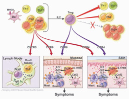
Patients with PA had tissue- and follicle-homing peanut-responsive CD4+ T cells with a heterogeneous pattern of TH2 differentiation, whereas control subjects had undetectable T-cell responses to peanut. The PA group had a delayed and IL-2-dependent upregulation of CD154 on cells expressing regulatory T (Treg) cell markers, which was absent in HC or high-threshold subjects. Depletion of Treg cells enhanced cytokine production in HC subjects and patients with PA in vitro, but cytokines associated with highly differentiated TH2 cells were more resistant to Treg cell suppression in patients with PA. Analysis of gene expression by means of single-cell RNA sequencing identified T cells with highly correlated expression of IL4, IL5, IL9, IL13, and the IL-25 receptor IL17RB.
CONCLUSIONS
These results demonstrate the presence of highly differentiated TH2 cells producing TH2-associated cytokines with functions beyond IgE class-switching in patients with PA. A multifunctional TH2 response was more evident than a Treg cell deficit among peanut-responsive T cells.
中文翻译:

花生过敏患者中花生反应性T细胞的单细胞分析揭示了异源效应子TH2亚型。
背景技术花生特异性T细胞的表型变异对临床过敏或对花生的耐受性的贡献尚不清楚。目的我们的目标是对患有或不患有花生过敏(PA)的受试者外周血中的花生特异性T细胞进行全面表型分析。方法我们从PA患者中获得了样本,其中包括接受基线花生攻击的队列进行免疫治疗试验(食品过敏研究联盟[CoFAR] 6)。受试者被确认患有PA,或者如果他们通过了1-g花生激发试验,则被称为高阈值受试者。还招募了健康对照(HC)受试者。在用花生提取物刺激6至18小时后,根据CD154的表达鉴定出花生反应性T细胞。使用流式细胞仪和单细胞RNA测序分析细胞。结果PA患者的组织和卵泡归巢的花生反应性CD4 + T细胞具有TH2分化的异质模式,而对照组则无法检测到对花生的T细胞反应。PA组在表达调节性T(Treg)细胞标志物的细胞上CD154具有延迟的和依赖IL-2的上调表达,而HC或高阈值受试者中则不存在。Treg细胞的耗竭在体外增强了HC受试者和PA患者的细胞因子产生,但是与高分化TH2细胞相关的细胞因子对PA患者的Treg细胞抑制作用更具有抵抗力。通过单细胞RNA测序对基因表达进行分析,鉴定出与IL4,IL5,IL9,IL13和IL-25受体IL17RB高度相关的T细胞。结论这些结果表明,在PA患者中,存在高度分化的TH2细胞,该细胞产生TH2相关细胞因子,其功能超出IgE类转换。在花生反应性T细胞中,多功能TH2反应比Treg细胞缺乏症更为明显。
更新日期:2018-01-31
中文翻译:

花生过敏患者中花生反应性T细胞的单细胞分析揭示了异源效应子TH2亚型。
背景技术花生特异性T细胞的表型变异对临床过敏或对花生的耐受性的贡献尚不清楚。目的我们的目标是对患有或不患有花生过敏(PA)的受试者外周血中的花生特异性T细胞进行全面表型分析。方法我们从PA患者中获得了样本,其中包括接受基线花生攻击的队列进行免疫治疗试验(食品过敏研究联盟[CoFAR] 6)。受试者被确认患有PA,或者如果他们通过了1-g花生激发试验,则被称为高阈值受试者。还招募了健康对照(HC)受试者。在用花生提取物刺激6至18小时后,根据CD154的表达鉴定出花生反应性T细胞。使用流式细胞仪和单细胞RNA测序分析细胞。结果PA患者的组织和卵泡归巢的花生反应性CD4 + T细胞具有TH2分化的异质模式,而对照组则无法检测到对花生的T细胞反应。PA组在表达调节性T(Treg)细胞标志物的细胞上CD154具有延迟的和依赖IL-2的上调表达,而HC或高阈值受试者中则不存在。Treg细胞的耗竭在体外增强了HC受试者和PA患者的细胞因子产生,但是与高分化TH2细胞相关的细胞因子对PA患者的Treg细胞抑制作用更具有抵抗力。通过单细胞RNA测序对基因表达进行分析,鉴定出与IL4,IL5,IL9,IL13和IL-25受体IL17RB高度相关的T细胞。结论这些结果表明,在PA患者中,存在高度分化的TH2细胞,该细胞产生TH2相关细胞因子,其功能超出IgE类转换。在花生反应性T细胞中,多功能TH2反应比Treg细胞缺乏症更为明显。


 京公网安备 11010802027423号