当前位置:
X-MOL 学术
›
J. Anal. At. Spectrom.
›
论文详情
Our official English website, www.x-mol.net, welcomes your feedback! (Note: you will need to create a separate account there.)
Combination of micro X-ray fluorescence spectroscopy and time-of-flight secondary ion mass spectrometry imaging for the marker-free detection of CeO2 nanoparticles in tissue sections†
Journal of Analytical Atomic Spectrometry ( IF 3.4 ) Pub Date : 2018-01-30 00:00:00 , DOI: 10.1039/c7ja00325k Lothar Veith 1, 2, 3, 4, 5 , Dörthe Dietrich 2, 3, 6, 7 , Antje Vennemann 2, 3, 8 , Daniel Breitenstein 1, 2, 3 , Carsten Engelhard 3, 4, 5, 9 , Uwe Karst 2, 3, 6, 7 , Michael Sperling 2, 3, 6, 7 , Martin Wiemann 2, 3, 8 , Birgit Hagenhoff 1, 2, 3
Journal of Analytical Atomic Spectrometry ( IF 3.4 ) Pub Date : 2018-01-30 00:00:00 , DOI: 10.1039/c7ja00325k Lothar Veith 1, 2, 3, 4, 5 , Dörthe Dietrich 2, 3, 6, 7 , Antje Vennemann 2, 3, 8 , Daniel Breitenstein 1, 2, 3 , Carsten Engelhard 3, 4, 5, 9 , Uwe Karst 2, 3, 6, 7 , Michael Sperling 2, 3, 6, 7 , Martin Wiemann 2, 3, 8 , Birgit Hagenhoff 1, 2, 3
Affiliation

|
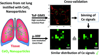
The description of nanoparticle distributions in tissue and associated effects is an important goal of nanotoxicology. Three-dimensional time-of-flight-secondary ion mass spectrometry (3D ToF-SIMS) is a promising label-free method to analyse tissue sections for nanoparticle distribution and to simultaneously detect organic and inorganic molecule species at high spatial resolution. As sample regions accessible to ToF-SIMS 3D imaging are limited to several hundred square micrometers, the proper selection of nanoparticle containing regions is mandatory. Micro X-ray fluorescence spectrometry (μ-XRF) provides a non-destructive, fast elemental analysis of large tissue sections with lateral resolution in the micrometer range. The latter technique was, therefore, used to screen samples as well as to validate elemental distribution images from ToF-SIMS 3D analysis. Specifically, cryo-sections from lungs inhomogeneously laden with CeO2 nanoparticles (10–200 nm) were subjected to μ-XRF and reflected light microscopy to pre-select lung tissue areas containing CeO2 NPs. The lateral resolution (670 nm) of ToF-SIMS imaging analyses in these areas exceeds the lateral resolution of μ-XRF imaging by a factor of 42 and reveals the lateral distribution of CeO2 NPs within the tissue structure. In addition, simultaneously acquired further secondary ions at a mass resolution of R = 4000 shed light on the sample composition. In summary, the investigation shows that μ-XRF is highly suitable to identify relevant tissue regions for subsequent high resolution ToF-SIMS 3D microanalysis of nanoparticle-laden tissue sections.
中文翻译:

显微X射线荧光光谱和飞行时间二次离子质谱成像技术的结合,用于组织切片中CeO 2纳米颗粒的无标记检测†
对组织中纳米颗粒分布及其相关作用的描述是纳米毒理学的重要目标。三维飞行时间二次离子质谱(3D ToF-SIMS)是一种很有前途的无标记方法,可用于分析组织切片的纳米颗粒分布并以高空间分辨率同时检测有机和无机分子种类。由于ToF-SIMS 3D成像可访问的样品区域被限制在数百平方微米,因此必须正确选择包含纳米颗粒的区域。微型X射线荧光光谱仪(μ-XRF)可对大型组织切片进行无损,快速的元素分析,其横向分辨率在微米范围内。因此,后一种技术是 用于筛选样品以及验证来自ToF-SIMS 3D分析的元素分布图像。具体来说,来自肺部的冷冻切片不均匀地充满了CeO对2个纳米颗粒(10–200 nm)进行μ-XRF和反射光显微镜检查,以预先选择含有CeO 2 NP的肺组织区域。在这些区域中,ToF-SIMS成像分析的横向分辨率(670 nm)比μ-XRF成像的横向分辨率高42倍,并且揭示了组织结构内CeO 2 NP的横向分布。另外,同时获得的质量分数为R = 4000的其他次级离子使样品组合物发光。总而言之,研究表明,μ-XRF非常适合识别相关组织区域,用于随后的纳米颗粒载有组织切片的高分辨率ToF-SIMS 3D微分析。
更新日期:2018-01-30
中文翻译:

显微X射线荧光光谱和飞行时间二次离子质谱成像技术的结合,用于组织切片中CeO 2纳米颗粒的无标记检测†
对组织中纳米颗粒分布及其相关作用的描述是纳米毒理学的重要目标。三维飞行时间二次离子质谱(3D ToF-SIMS)是一种很有前途的无标记方法,可用于分析组织切片的纳米颗粒分布并以高空间分辨率同时检测有机和无机分子种类。由于ToF-SIMS 3D成像可访问的样品区域被限制在数百平方微米,因此必须正确选择包含纳米颗粒的区域。微型X射线荧光光谱仪(μ-XRF)可对大型组织切片进行无损,快速的元素分析,其横向分辨率在微米范围内。因此,后一种技术是 用于筛选样品以及验证来自ToF-SIMS 3D分析的元素分布图像。具体来说,来自肺部的冷冻切片不均匀地充满了CeO对2个纳米颗粒(10–200 nm)进行μ-XRF和反射光显微镜检查,以预先选择含有CeO 2 NP的肺组织区域。在这些区域中,ToF-SIMS成像分析的横向分辨率(670 nm)比μ-XRF成像的横向分辨率高42倍,并且揭示了组织结构内CeO 2 NP的横向分布。另外,同时获得的质量分数为R = 4000的其他次级离子使样品组合物发光。总而言之,研究表明,μ-XRF非常适合识别相关组织区域,用于随后的纳米颗粒载有组织切片的高分辨率ToF-SIMS 3D微分析。


 京公网安备 11010802027423号