当前位置:
X-MOL 学术
›
ACS Chem. Neurosci.
›
论文详情
Our official English website, www.x-mol.net, welcomes your feedback! (Note: you will need to create a separate account there.)
Revealing the Penumbra through Imaging Elemental Markers of Cellular Metabolism in an Ischemic Stroke Model
ACS Chemical Neuroscience ( IF 5 ) Pub Date : 2018-01-25 00:00:00 , DOI: 10.1021/acschemneuro.7b00382 M. Jake Pushie 1 , Andrew M. Crawford 2 , Nicole J. Sylvain 1 , Huishu Hou 1 , Mark J. Hackett 3, 4 , Graham N. George 2 , Michael E. Kelly 1
ACS Chemical Neuroscience ( IF 5 ) Pub Date : 2018-01-25 00:00:00 , DOI: 10.1021/acschemneuro.7b00382 M. Jake Pushie 1 , Andrew M. Crawford 2 , Nicole J. Sylvain 1 , Huishu Hou 1 , Mark J. Hackett 3, 4 , Graham N. George 2 , Michael E. Kelly 1
Affiliation
|
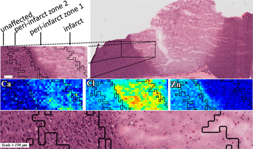
Stroke exacts a heavy financial and economic burden, is a leading cause of death, and is the leading cause of long-term disability in those who survive. The penumbra surrounds the ischemic core of the stroke lesion and is composed of cells that are stressed and vulnerable to death, which is due to an altered metabolic, oxidative, and ionic environment within the penumbra. Without therapeutic intervention, many cells within the penumbra will die and become part of the growing infarct, however, there is hope that appropriate therapies may allow potential recovery of cells within this tissue region, or at least slow the rate of cell death, therefore, slowing the spread of the ischemic infarct and minimizing the extent of tissue damage. As such, preserving the penumbra to promote functional brain recovery is a central goal in stroke research. While identification of the ischemic infarct, and the infarct/penumbra boundary is relatively trivial using classical histology and microscopy techniques, accurately assessing the penetration of the penumbra zone into undamaged brain tissue, and evaluating the magnitude of chemical alterations in the penumbra, has long been a major challenge to the stroke research field. In this study, we have used synchrotron-based X-ray fluorescence imaging to visualize the elemental changes in undamaged, penumbra, and infarct brain tissue, following ischemic stroke. We have employed a Gaussian mixture model to cluster tissue areas based on their elemental characteristics. The method separates the core of the infarct from healthy tissue, and also demarcates discrete regions encircling the infarct. These regions of interest can be combined with elemental and metabolic data, as well as with conventional histology. The cell populations defined by clustering provide a reproducible means of visualizing the size and extent of the penumbra at the level of the single cell and provide a critically needed tool to track changes in elemental status and penumbra size.
中文翻译:

在缺血性中风模型中通过成像细胞代谢的元素标志物揭示半影
中风造成沉重的经济和经济负担,是导致死亡的主要原因,也是导致生存者长期残疾的主要原因。半影围绕中风病灶的缺血核心,由受压且易于死亡的细胞组成,这是由于半影内新陈代谢,氧化和离子环境的改变所致。如果没有治疗干预,半影内的许多细胞将死亡并成为梗死灶的一部分,但是,人们希望适当的治疗方法可以使该组织区域内的细胞潜在地恢复,或者至少减慢细胞死亡的速度,因此,减缓缺血性梗塞的扩散并最大程度地减少组织损伤的程度。因此,保护半影以促进大脑功能恢复是中风研究的中心目标。虽然使用经典的组织学和显微镜技术识别缺血性梗塞和梗塞/半影边界相对比较琐碎,但准确评估半影带进入未受损脑组织的渗透率以及评估半影中化学变化的程度却一直很长。对卒中研究领域的重大挑战。在这项研究中,我们使用了基于同步加速器的X射线荧光成像技术,以观察缺血性中风后未受损,半影和梗死的脑组织中元素的变化。我们采用了高斯混合模型来根据其元素特征对组织区域进行聚类。该方法将梗塞的核心与健康组织分开,并且还划定了包围梗塞的离散区域。这些感兴趣的区域可以与元素和代谢数据以及常规组织学相结合。通过聚类定义的细胞群提供了一种可重现的方法,可以在单个细胞水平上可视化半影的大小和范围,并提供了跟踪元素状态和半影大小变化的迫切需要的工具。
更新日期:2018-01-25
中文翻译:
在缺血性中风模型中通过成像细胞代谢的元素标志物揭示半影
中风造成沉重的经济和经济负担,是导致死亡的主要原因,也是导致生存者长期残疾的主要原因。半影围绕中风病灶的缺血核心,由受压且易于死亡的细胞组成,这是由于半影内新陈代谢,氧化和离子环境的改变所致。如果没有治疗干预,半影内的许多细胞将死亡并成为梗死灶的一部分,但是,人们希望适当的治疗方法可以使该组织区域内的细胞潜在地恢复,或者至少减慢细胞死亡的速度,因此,减缓缺血性梗塞的扩散并最大程度地减少组织损伤的程度。因此,保护半影以促进大脑功能恢复是中风研究的中心目标。虽然使用经典的组织学和显微镜技术识别缺血性梗塞和梗塞/半影边界相对比较琐碎,但准确评估半影带进入未受损脑组织的渗透率以及评估半影中化学变化的程度却一直很长。对卒中研究领域的重大挑战。在这项研究中,我们使用了基于同步加速器的X射线荧光成像技术,以观察缺血性中风后未受损,半影和梗死的脑组织中元素的变化。我们采用了高斯混合模型来根据其元素特征对组织区域进行聚类。该方法将梗塞的核心与健康组织分开,并且还划定了包围梗塞的离散区域。这些感兴趣的区域可以与元素和代谢数据以及常规组织学相结合。通过聚类定义的细胞群提供了一种可重现的方法,可以在单个细胞水平上可视化半影的大小和范围,并提供了跟踪元素状态和半影大小变化的迫切需要的工具。


 京公网安备 11010802027423号