当前位置:
X-MOL 学术
›
Adv. Funct. Mater.
›
论文详情
Our official English website, www.x-mol.net, welcomes your feedback! (Note: you will need to create a separate account there.)
Polydimethylsiloxane Composites for Optical Ultrasound Generation and Multimodality Imaging
Advanced Functional Materials ( IF 19.0 ) Pub Date : 2018-01-15 , DOI: 10.1002/adfm.201704919 Sacha Noimark 1, 2, 3 , Richard J. Colchester 1, 2 , Radhika K. Poduval 4 , Efthymios Maneas 1, 2 , Erwin J. Alles 1, 2 , Tianrui Zhao 3 , Edward Z. Zhang 1, 2 , Michael Ashworth 5 , Elena Tsolaki 2 , Adrian H. Chester 6 , Najma Latif 6 , Sergio Bertazzo 2 , Anna L. David 1, 7 , Sebastien Ourselin 1, 2 , Paul C. Beard 1, 2 , Ivan P. Parkin 1, 3 , Ioannis Papakonstantinou 4 , Adrien E. Desjardins 1, 2
Advanced Functional Materials ( IF 19.0 ) Pub Date : 2018-01-15 , DOI: 10.1002/adfm.201704919 Sacha Noimark 1, 2, 3 , Richard J. Colchester 1, 2 , Radhika K. Poduval 4 , Efthymios Maneas 1, 2 , Erwin J. Alles 1, 2 , Tianrui Zhao 3 , Edward Z. Zhang 1, 2 , Michael Ashworth 5 , Elena Tsolaki 2 , Adrian H. Chester 6 , Najma Latif 6 , Sergio Bertazzo 2 , Anna L. David 1, 7 , Sebastien Ourselin 1, 2 , Paul C. Beard 1, 2 , Ivan P. Parkin 1, 3 , Ioannis Papakonstantinou 4 , Adrien E. Desjardins 1, 2
Affiliation
|
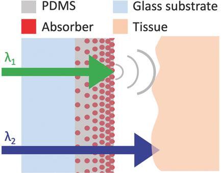
Polydimethylsiloxane (PDMS) is widely used in biomedical science and can form composites that have broad applicability. One promising application where PDMS composites offer several advantages is optical ultrasound generation via the photoacoustic effect. Here, methods to create these PDMS composites are reviewed and classified. It is highlighted how the composites can be applied to a range of substrates, from micrometer‐scale, temperature‐sensitive optical fibers to centimeter‐scale curved and planar surfaces. The resulting composites have enabled all‐optical ultrasound imaging of biological tissues both ex vivo and in vivo, with high spatial resolution and with clinically relevant contrast. In addition, the first 3D all‐optical pulse‐echo ultrasound imaging of ex vivo human tissue, using a PDMS‐multiwalled carbon nanotube composite and a fiber‐optic ultrasound receiver, is presented. Gold nanoparticle‐PDMS and crystal violet‐PDMS composites with prominent absorption at one wavelength range for pulse‐echo ultrasound imaging and transmission at a second wavelength range for photoacoustic imaging are also presented. Using these devices, images of diseased human vascular tissue with both structural and molecular contrast are obtained. With a broader perspective, literature on recent advances in PDMS microfabrication from different fields is highlighted, and methods for incorporating them into new generations of optical ultrasound generators are suggested.
中文翻译:

用于光学超声产生和多峰成像的聚二甲基硅氧烷复合材料
聚二甲基硅氧烷(PDMS)在生物医学领域被广泛使用,并且可以形成具有广泛适用性的复合材料。PDMS复合材料具有多种优势的一项有前途的应用是通过光声效应产生光学超声。在这里,对这些PDMS复合材料的制造方法进行了回顾和分类。重点介绍了如何将复合材料应用于各种基材,从微米级,对温度敏感的光纤到厘米级弯曲和平面表面。由此产生的复合材料能够实现离体和体内生物组织的全光学超声成像,具有高空间分辨率和临床相关的对比度。此外,首次对离体人体组织进行3D全光脉冲回波超声成像,介绍了使用PDMS多壁碳纳米管复合材料和光纤超声接收器的方法。还介绍了金纳米颗粒PDMS和结晶紫PDMS复合材料,它们在一个波长范围内具有明显的吸收性,可用于脉冲回波超声成像,而在另一波长范围内具有透射性,可用于光声成像。使用这些设备,可以获得具有结构和分子对比的病变人类血管组织的图像。从更广阔的角度来看,着重介绍了不同领域PDMS微制造的最新进展,并提出了将其纳入新一代光学超声发生器的方法。还介绍了金纳米颗粒PDMS和结晶紫PDMS复合材料,它们在一个波长范围内具有明显的吸收性,可用于脉冲回波超声成像,而在另一波长范围内具有透射性,可用于光声成像。使用这些设备,可以获得具有结构和分子对比的病变人类血管组织的图像。从更广阔的角度来看,着重介绍了不同领域PDMS微制造的最新进展,并提出了将其纳入新一代光学超声发生器的方法。还介绍了金纳米颗粒PDMS和结晶紫PDMS复合材料,它们在一个波长范围内具有明显的吸收性,可用于脉冲回波超声成像,而在另一波长范围内具有透射性,可用于光声成像。使用这些设备,可以获得具有结构和分子对比的病变人类血管组织的图像。从更广阔的角度来看,着重介绍了不同领域PDMS微制造的最新进展,并提出了将其纳入新一代光学超声发生器的方法。
更新日期:2018-01-15
中文翻译:
用于光学超声产生和多峰成像的聚二甲基硅氧烷复合材料
聚二甲基硅氧烷(PDMS)在生物医学领域被广泛使用,并且可以形成具有广泛适用性的复合材料。PDMS复合材料具有多种优势的一项有前途的应用是通过光声效应产生光学超声。在这里,对这些PDMS复合材料的制造方法进行了回顾和分类。重点介绍了如何将复合材料应用于各种基材,从微米级,对温度敏感的光纤到厘米级弯曲和平面表面。由此产生的复合材料能够实现离体和体内生物组织的全光学超声成像,具有高空间分辨率和临床相关的对比度。此外,首次对离体人体组织进行3D全光脉冲回波超声成像,介绍了使用PDMS多壁碳纳米管复合材料和光纤超声接收器的方法。还介绍了金纳米颗粒PDMS和结晶紫PDMS复合材料,它们在一个波长范围内具有明显的吸收性,可用于脉冲回波超声成像,而在另一波长范围内具有透射性,可用于光声成像。使用这些设备,可以获得具有结构和分子对比的病变人类血管组织的图像。从更广阔的角度来看,着重介绍了不同领域PDMS微制造的最新进展,并提出了将其纳入新一代光学超声发生器的方法。还介绍了金纳米颗粒PDMS和结晶紫PDMS复合材料,它们在一个波长范围内具有明显的吸收性,可用于脉冲回波超声成像,而在另一波长范围内具有透射性,可用于光声成像。使用这些设备,可以获得具有结构和分子对比的病变人类血管组织的图像。从更广阔的角度来看,着重介绍了不同领域PDMS微制造的最新进展,并提出了将其纳入新一代光学超声发生器的方法。还介绍了金纳米颗粒PDMS和结晶紫PDMS复合材料,它们在一个波长范围内具有明显的吸收性,可用于脉冲回波超声成像,而在另一波长范围内具有透射性,可用于光声成像。使用这些设备,可以获得具有结构和分子对比的病变人类血管组织的图像。从更广阔的角度来看,着重介绍了不同领域PDMS微制造的最新进展,并提出了将其纳入新一代光学超声发生器的方法。


 京公网安备 11010802027423号