Our official English website, www.x-mol.net, welcomes your feedback! (Note: you will need to create a separate account there.)
Cycloid scanning for wide field optical coherence tomography endomicroscopy and angiography in vivo
Optica ( IF 10.4 ) Pub Date : 2018-01-11 , DOI: 10.1364/optica.5.000036 Kaicheng Liang , Zhao Wang , Osman O. Ahsen , Hsiang-Chieh Lee , Benjamin M. Potsaid , Vijaysekhar Jayaraman , Alex Cable , Hiroshi Mashimo , Xingde Li , James G. Fujimoto
Optica ( IF 10.4 ) Pub Date : 2018-01-11 , DOI: 10.1364/optica.5.000036 Kaicheng Liang , Zhao Wang , Osman O. Ahsen , Hsiang-Chieh Lee , Benjamin M. Potsaid , Vijaysekhar Jayaraman , Alex Cable , Hiroshi Mashimo , Xingde Li , James G. Fujimoto

|
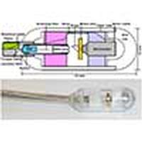
Devices that perform wide field-of-view (FOV) precision optical scanning are important for endoscopic assessment and diagnosis of luminal organ disease such as in gastroenterology. Optical scanning for in vivo endoscopic imaging has traditionally relied on one or more proximal mechanical actuators, limiting scan accuracy and imaging speed. There is a need for rapid and precise two-dimensional (2D) microscanning technologies to enable the translation of benchtop scanning microscopies to in vivo endoscopic imaging. We demonstrate a new cycloid scanner in a tethered capsule for ultrahigh speed, side-viewing optical coherence tomography (OCT) endomicroscopy in vivo. The cycloid capsule incorporates two scanners: a piezoelectrically actuated resonant fiber scanner to perform a precision, small FOV, fast scan and a micromotor scanner to perform a wide FOV, slow scan. Together these scanners distally scan the beam circumferentially in a 2D cycloid pattern, generating an unwrapped strip FOV. Sequential strip volumes can be acquired with proximal pullback to image centimeter-long regions. Using ultrahigh speed 1.3 μm wavelength swept-source OCT at a 1.17 MHz axial scan rate, we imaged the human rectum at 3 volumes/s. Each OCT strip volume had axial scans with 8.5 μm axial and 30 μm transverse resolution. We further demonstrate OCT angiography at 0.5 volumes/s, producing volumetric images of vasculature. In addition to OCT applications, cycloid scanning promises to enable precision 2D optical scanning for other imaging modalities, including fluorescence confocal and nonlinear microscopy.
中文翻译:

摆线扫描在体内进行广域光学相干断层扫描内窥镜和血管造影
执行宽视场(FOV)精密光学扫描的设备对于内窥镜评估和诊断腔内器官疾病(例如胃肠病学)非常重要。用于体内内窥镜成像的光学扫描传统上依赖于一个或多个近端机械致动器,从而限制了扫描精度和成像速度。需要快速和精确的二维(2D)显微扫描技术,以将台式扫描显微图像转换为体内内窥镜成像。我们展示了一种栓状胶囊中的新型摆线扫描仪,用于体内超高速,侧视光学相干断层扫描(OCT)内窥镜检查。摆线针囊包含两个扫描仪:一个压电致动的共振光纤扫描仪,用于执行精密的小FOV快速扫描;以及微电机扫描仪,用于执行宽的FOV,慢速扫描。这些扫描仪一起以2D摆线模式向远侧扫描光束,生成未包裹的光束带状视场。可以通过将近端拉回至图像厘米长的区域来获取连续的条带体积。使用1.17 MHz轴向扫描速率的超高速1.3μm波长扫频源OCT,我们以3次/ s的速度对人体直肠成像。每个OCT带材体积轴向扫描,轴向分辨率为8.5μm,横向分辨率为30μm。我们进一步证明了OCT血管造影术在0.5卷/ s,产生血管的体积图像。除OCT应用外,摆线扫描有望为其他成像方式(包括荧光共聚焦和非线性显微镜)实现精确的2D光学扫描。
更新日期:2018-01-19
中文翻译:

摆线扫描在体内进行广域光学相干断层扫描内窥镜和血管造影
执行宽视场(FOV)精密光学扫描的设备对于内窥镜评估和诊断腔内器官疾病(例如胃肠病学)非常重要。用于体内内窥镜成像的光学扫描传统上依赖于一个或多个近端机械致动器,从而限制了扫描精度和成像速度。需要快速和精确的二维(2D)显微扫描技术,以将台式扫描显微图像转换为体内内窥镜成像。我们展示了一种栓状胶囊中的新型摆线扫描仪,用于体内超高速,侧视光学相干断层扫描(OCT)内窥镜检查。摆线针囊包含两个扫描仪:一个压电致动的共振光纤扫描仪,用于执行精密的小FOV快速扫描;以及微电机扫描仪,用于执行宽的FOV,慢速扫描。这些扫描仪一起以2D摆线模式向远侧扫描光束,生成未包裹的光束带状视场。可以通过将近端拉回至图像厘米长的区域来获取连续的条带体积。使用1.17 MHz轴向扫描速率的超高速1.3μm波长扫频源OCT,我们以3次/ s的速度对人体直肠成像。每个OCT带材体积轴向扫描,轴向分辨率为8.5μm,横向分辨率为30μm。我们进一步证明了OCT血管造影术在0.5卷/ s,产生血管的体积图像。除OCT应用外,摆线扫描有望为其他成像方式(包括荧光共聚焦和非线性显微镜)实现精确的2D光学扫描。


 京公网安备 11010802027423号