当前位置:
X-MOL 学术
›
Anal. Methods
›
论文详情
Our official English website, www.x-mol.net, welcomes your feedback! (Note: you will need to create a separate account there.)
Quantitative imaging of translocated silver following nanoparticle exposure by laser ablation-inductively coupled plasma-mass spectrometry
Analytical Methods ( IF 3.1 ) Pub Date : 2018-01-10 00:00:00 , DOI: 10.1039/c7ay02294h David P. Bishop 1, 2, 3, 4 , Mandy Grossgarten 5, 6, 7, 8 , Dörthe Dietrich 5, 6, 7, 8 , Antje Vennemann 7, 8, 9 , Nerida Cole 1, 2, 3, 4 , Michael Sperling 5, 6, 7, 8 , Martin Wiemann 7, 8, 9 , Philip A. Doble 1, 2, 3, 4 , Uwe Karst 5, 6, 7, 8
Analytical Methods ( IF 3.1 ) Pub Date : 2018-01-10 00:00:00 , DOI: 10.1039/c7ay02294h David P. Bishop 1, 2, 3, 4 , Mandy Grossgarten 5, 6, 7, 8 , Dörthe Dietrich 5, 6, 7, 8 , Antje Vennemann 7, 8, 9 , Nerida Cole 1, 2, 3, 4 , Michael Sperling 5, 6, 7, 8 , Martin Wiemann 7, 8, 9 , Philip A. Doble 1, 2, 3, 4 , Uwe Karst 5, 6, 7, 8
Affiliation
|
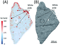
The likelihood of exposure to antimicrobial silver nanoparticles continues to grow with their increasing ubiquity in various medical and consumer products. While translocation of silver nanoparticles to major organs has been examined, the in situ location and concentration in the organs is not well characterised. Here we have used laser ablation-inductively coupled plasma-mass spectrometry to quantitatively image serial sections to construct a three-dimensional representation of the distribution of silver in rat spleen following respiratory tract exposure via intratracheal instillation of silver nanoparticles. Silver was distributed predominantly in the white pulp of the spleen at concentrations greater than 300 ng g−1. Imaging tissue sections via laser-ablation-inductively coupled plasma-mass spectrometry is an excellent tool for the visualisation and quantification of metals attributed to nanoparticles in organs allowing investigation of silver nanoparticle exposure in vivo.
中文翻译:

激光烧蚀-电感耦合等离子体质谱法对纳米粒子暴露后的易位银进行定量成像
随着它们在各种医疗和消费产品中的普及,暴露于抗菌银纳米颗粒的可能性持续增长。虽然已经检查了银纳米颗粒向主要器官的转运,但是器官中的原位定位和浓度并未得到很好的表征。在这里,我们已经使用激光烧蚀-电感耦合等离子体质谱法对连续切片进行定量成像,以构建三维三维表示大鼠气管内通过气管内滴注银纳米颗粒暴露于呼吸道后的脾脏中银的分布情况。银主要以大于300 ng g -1的浓度分布在脾脏的白色牙髓中。成像组织切片通过激光烧蚀-电感耦合等离子体质谱法是一种出色的工具,可用于可视化和量化器官中属于纳米颗粒的金属,从而可以研究体内银纳米颗粒的暴露情况。
更新日期:2018-01-10
中文翻译:
激光烧蚀-电感耦合等离子体质谱法对纳米粒子暴露后的易位银进行定量成像
随着它们在各种医疗和消费产品中的普及,暴露于抗菌银纳米颗粒的可能性持续增长。虽然已经检查了银纳米颗粒向主要器官的转运,但是器官中的原位定位和浓度并未得到很好的表征。在这里,我们已经使用激光烧蚀-电感耦合等离子体质谱法对连续切片进行定量成像,以构建三维三维表示大鼠气管内通过气管内滴注银纳米颗粒暴露于呼吸道后的脾脏中银的分布情况。银主要以大于300 ng g -1的浓度分布在脾脏的白色牙髓中。成像组织切片通过激光烧蚀-电感耦合等离子体质谱法是一种出色的工具,可用于可视化和量化器官中属于纳米颗粒的金属,从而可以研究体内银纳米颗粒的暴露情况。


 京公网安备 11010802027423号