Our official English website, www.x-mol.net, welcomes your feedback! (Note: you will need to create a separate account there.)
Monitoring the dynamics of cell-derived extracellular vesicles at the nanoscale by liquid-cell transmission electron microscopy†
Nanoscale ( IF 6.7 ) Pub Date : 2018-01-02 00:00:00 , DOI: 10.1039/c7nr07576f Max Piffoux 1, 2, 3, 4, 5 , Nabeel Ahmad 3, 4, 6, 7 , Jaysen Nelayah 3, 4, 6, 7 , Claire Wilhelm 1, 2, 3, 4 , Amanda Silva 1, 2, 3, 4 , Florence Gazeau 1, 2, 3, 4 , Damien Alloyeau 3, 4, 6, 7
Nanoscale ( IF 6.7 ) Pub Date : 2018-01-02 00:00:00 , DOI: 10.1039/c7nr07576f Max Piffoux 1, 2, 3, 4, 5 , Nabeel Ahmad 3, 4, 6, 7 , Jaysen Nelayah 3, 4, 6, 7 , Claire Wilhelm 1, 2, 3, 4 , Amanda Silva 1, 2, 3, 4 , Florence Gazeau 1, 2, 3, 4 , Damien Alloyeau 3, 4, 6, 7
Affiliation
|
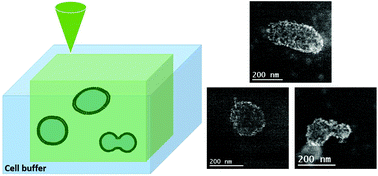
Cell-derived extracellular vesicles (EVs) circulating in body fluids hold promises as bioactive therapeutic agents and as biomarkers to diagnose a wide range of diseases. However nano-imaging methods are needed to characterize these complex and heterogeneous soft materials in their native wet environment. Herein, we exploit liquid-cell transmission electron microscopy (LCTEM) to characterize the morphology and dynamic behavior of EVs in physiological media with nanometer resolution. The beam-induced controlled growth of Au nanoparticles on bilayer membranes is used as an original in situ staining method to improve the contrast of EVs and artificial liposomes. LCTEM provides information about the size distribution and concentration of EVs that are consistent with Cryo-TEM and nanoparticle tracking analysis measurements. Moreover, LCTEM gives a unique insight into the dynamics of EVs depending on their liquid environment. The size-dependent morphology of EVs is sensitive to osmotic stress which tends to transform their spherical shape to ellipsoidal, stomatocyte or discocyte morphologies. In the liquid-cell, EVs exhibit a sub-diffusive motion due to strong interactions between the Au nanoparticles and the liquid-cell windows. Finally, the high-resolution monitoring of EV aggregation and fusion illustrate that LCTEM opens up a new way to study cell-membrane dynamics.
中文翻译:

通过液细胞透射电子显微镜在纳米尺度上监测细胞衍生的细胞外囊泡的动力学†
在体液中循环的源自细胞的细胞外囊泡(EV)具有作为生物活性治疗剂和诊断广泛疾病的生物标记物的希望。然而,需要纳米成像方法来表征这些复杂且异质的软质材料在其天然的潮湿环境中。在这里,我们利用液体细胞透射电子显微镜(LCTEM)来表征具有纳米分辨率的生理介质中电动汽车的形态和动态行为。光束诱导的金纳米颗粒在双层膜上的受控生长被用作原始的原位染色方法可提高电动汽车与人工脂质体的对比度。LCTEM提供有关EV的尺寸分布和浓度的信息,这些信息与Cryo-TEM和纳米颗粒跟踪分析测量结果一致。此外,LCTEM可以根据电动汽车的液体环境对电动汽车的动力学特性提供独特的见解。电动汽车的尺寸依赖性形态对渗透压敏感,渗透压趋于将其球形转变为椭圆形,口腔细胞或盘状细胞形态。在液池中,由于Au纳米颗粒与液池窗口之间的强相互作用,因此EV表现出亚扩散运动。最后,对EV聚集和融合的高分辨率监测表明,LCTEM开辟了一种研究细胞膜动力学的新方法。
更新日期:2018-01-02
中文翻译:
通过液细胞透射电子显微镜在纳米尺度上监测细胞衍生的细胞外囊泡的动力学†
在体液中循环的源自细胞的细胞外囊泡(EV)具有作为生物活性治疗剂和诊断广泛疾病的生物标记物的希望。然而,需要纳米成像方法来表征这些复杂且异质的软质材料在其天然的潮湿环境中。在这里,我们利用液体细胞透射电子显微镜(LCTEM)来表征具有纳米分辨率的生理介质中电动汽车的形态和动态行为。光束诱导的金纳米颗粒在双层膜上的受控生长被用作原始的原位染色方法可提高电动汽车与人工脂质体的对比度。LCTEM提供有关EV的尺寸分布和浓度的信息,这些信息与Cryo-TEM和纳米颗粒跟踪分析测量结果一致。此外,LCTEM可以根据电动汽车的液体环境对电动汽车的动力学特性提供独特的见解。电动汽车的尺寸依赖性形态对渗透压敏感,渗透压趋于将其球形转变为椭圆形,口腔细胞或盘状细胞形态。在液池中,由于Au纳米颗粒与液池窗口之间的强相互作用,因此EV表现出亚扩散运动。最后,对EV聚集和融合的高分辨率监测表明,LCTEM开辟了一种研究细胞膜动力学的新方法。


 京公网安备 11010802027423号