Journal of the Mechanical Behavior of Biomedical Materials ( IF 3.9 ) Pub Date : 2017-12-16 , DOI: 10.1016/j.jmbbm.2017.12.015 Peng-Fei Yang , Xiao-Tong Nie , Dong-Dong Zhao , Zhe Wang , Li Ren , Hui-Yun Xu , Joern Rittweger , Peng Shang
|
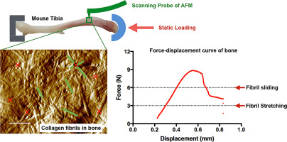
The mechanical properties of the bone play a decisive role in the resistance of the bone to fracture. Clinically, the quantity of the bone in the mineral phase has been considered as the gold-standard indicator for the risk of bone fracture. However, the bone is a complex tissue with a hierarchical-structure consisting of organic matrix, mineral hydroxyapatite, and water. Collagen comprises up to 90% of the organic matrix in the bone, and is vital for its mechanical behavior. To date, the morphological and mechanical responses of collagen fibrils in the bone matrix have been largely overlooked. In the present study, an atomic force microscopy-based imaging and indentation approach is introduced and integrated with a tibia axial loading model. The morphology of mineralized Type I collagen fibrils of the murine cortical tibia is imaged after demineralization, and the in situ elastic modulus of the fibrils is quantified at different loading conditions. Results suggested that the mineralized collagen fibrils are stretched in the early phase of bone deformation, characterized by the elongation of the D-periodic spacing. Reorientation of the collagen fibrils is demonstrated in the subsequent phase of bone deformation. The in situ radial elastic modulus of the collagen fibrils remained constant under the tested loading conditions. These experimental findings provide evidence in support of the unique deformation regimes of bone tissue from the perspective of alterations of mineralized collagen fibrils. This study allows the understanding of the unique mechanical behavior of the bone at the nanoscale, and reveals the mechanisms of relevant diseases that impair the mechanical properties of the bone.
中文翻译:
在机械载荷下通过原位形态和弹性模量观察揭示了皮质骨中胶原原纤维的变形方式
骨骼的机械性能在骨骼的抗断裂性中起着决定性的作用。临床上,矿物质相中的骨量已被视为骨折风险的金标准指标。但是,骨骼是复杂的组织,具有由有机基质,矿物羟基磷灰石和水组成的层次结构。胶原蛋白占骨骼中有机基质的90%,对于其机械行为至关重要。迄今为止,骨基质中胶原蛋白原纤维的形态和机械反应已被大大忽略。在本研究中,介绍了一种基于原子力显微镜的成像和压痕方法,并将其与胫骨轴向载荷模型集成在一起。在不同的负载条件下,原纤维的原位弹性模量被量化。结果表明矿化的胶原原纤维在骨变形的早期被拉伸,其特征在于D周期间隔的延长。在骨变形的随后阶段证明了胶原蛋白原纤维的重新定向。在原位在测试的负载条件下,胶原原纤维的径向弹性模量保持恒定。这些实验结果为矿化胶原蛋白原纤维的改变提供了支持骨组织独特变形机制的证据。这项研究可以了解纳米级骨骼的独特机械行为,并揭示损害骨骼机械性能的相关疾病的机制。


 京公网安备 11010802027423号