Ophthalmology ( IF 13.7 ) Pub Date : 2017-12-08 , DOI: 10.1016/j.ophtha.2017.10.031 Thomas Schlegl , Sebastian M. Waldstein , Hrvoje Bogunovic , Franz Endstraßer , Amir Sadeghipour , Ana-Maria Philip , Dominika Podkowinski , Bianca S. Gerendas , Georg Langs , Ursula Schmidt-Erfurth
|
Purpose
Development and validation of a fully automated method to detect and quantify macular fluid in conventional OCT images.
Design
Development of a diagnostic modality.
Participants
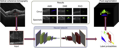
The clinical dataset for fluid detection consisted of 1200 OCT volumes of patients with neovascular age-related macular degeneration (AMD, n = 400), diabetic macular edema (DME, n = 400), or retinal vein occlusion (RVO, n = 400) acquired with Zeiss Cirrus (Carl Zeiss Meditec, Dublin, CA) (n = 600) or Heidelberg Spectralis (Heidelberg Engineering, Heidelberg, Germany) (n = 600) OCT devices.
Methods
A method based on deep learning to automatically detect and quantify intraretinal cystoid fluid (IRC) and subretinal fluid (SRF) was developed. The performance of the algorithm in accurately identifying fluid localization and extent was evaluated against a manual consensus reading of 2 masked reading center graders.
Main Outcome Measures
Performance of a fully automated method to accurately detect, differentiate, and quantify intraretinal and SRF using area under the receiver operating characteristics curves, precision, and recall.
Results
The newly designed, fully automated diagnostic method based on deep learning achieved optimal accuracy for the detection and quantification of IRC for all 3 macular pathologies with a mean accuracy (AUC) of 0.94 (range, 0.91–0.97), a mean precision of 0.91, and a mean recall of 0.84. The detection and measurement of SRF were also highly accurate with an AUC of 0.92 (range, 0.86–0.98), a mean precision of 0.61, and a mean recall of 0.81, with superior performance in neovascular AMD and RVO compared with DME, which was represented rarely in the population studied. High linear correlation was confirmed between automated and manual fluid localization and quantification, yielding an average Pearson's correlation coefficient of 0.90 for IRC and of 0.96 for SRF.
Conclusions
Deep learning in retinal image analysis achieves excellent accuracy for the differential detection of retinal fluid types across the most prevalent exudative macular diseases and OCT devices. Furthermore, quantification of fluid achieves a high level of concordance with manual expert assessment. Fully automated analysis of retinal OCT images from clinical routine provides a promising horizon in improving accuracy and reliability of retinal diagnosis for research and clinical practice in ophthalmology.
中文翻译:
使用深度学习对OCT中的黄斑液进行全自动检测和定量
目的
开发并验证了一种用于检测和定量常规OCT图像中的黄斑液的全自动方法。
设计
诊断方式的发展。
参加者
用于体液检测的临床数据集由1200例OCT体积的新生血管性年龄相关性黄斑变性(AMD,n = 400),糖尿病性黄斑水肿(DME,n = 400)或视网膜静脉阻塞(RVO,n = 400)的患者组成使用Zeiss Cirrus(Carl Zeiss Meditec,都柏林,CA)(n = 600)或Heidelberg Spectralis(Heidelberg Engineering,Heidelberg,Germany)(n = 600)OCT设备获得。
方法
开发了一种基于深度学习的方法,可以自动检测和定量视网膜内囊样液(IRC)和视网膜下液(SRF)。该算法在准确识别流体定位和范围方面的性能是根据2个带遮罩的阅读中心评分员的人工共识读数进行评估的。
主要观察指标
使用接收器工作特性曲线,准确度和召回率下的面积来精确检测,区分和量化视网膜内和SRF的全自动方法的性能。
结果
新设计的基于深度学习的全自动诊断方法在3种黄斑病变中对IRC的检测和定量均达到了最佳准确度,其平均准确度(AUC)为0.94(范围为0.91-0.97),平均准确度为0.91,平均召回率为0.84。SRF的检测和测量也非常准确,AUC为0.92(范围为0.86-0.98),平均精度为0.61,平均召回率为0.81,与DME相比,在新血管性AMD和RVO中的性能更好。在所研究的人群中很少代表。确认了自动和手动流体定位与定量之间的高线性相关性,IRC的平均皮尔逊相关系数为0.90,SRF的平均Pearson相关系数为0.96。
结论
在大多数流行的渗出性黄斑疾病和OCT设备中,视网膜图像分析的深度学习可实现出色的视网膜液类型差异检测精度。此外,流体的定量与手动专家评估达到了高度的一致性。从临床常规中对视网膜OCT图像进行全自动分析,为提高眼科研究和临床实践中视网膜诊断的准确性和可靠性提供了广阔的前景。


 京公网安备 11010802027423号