当前位置:
X-MOL 学术
›
Adv. Funct. Mater.
›
论文详情
Our official English website, www.x-mol.net, welcomes your feedback! (Note: you will need to create a separate account there.)
Photoacoustic Imaging of Embryonic Stem Cell‐Derived Cardiomyocytes in Living Hearts with Ultrasensitive Semiconducting Polymer Nanoparticles
Advanced Functional Materials ( IF 19.0 ) Pub Date : 2017-11-08 , DOI: 10.1002/adfm.201704939 Xulei Qin 1 , Haodong Chen 1 , Huaxiao Yang 1 , Haodi Wu 1 , Xin Zhao 1 , Huiyuan Wang 2 , Tony Chour 1 , Evgenios Neofytou 1 , Dan Ding 3 , Heike Daldrup-Link 4 , Sarah C Heilshorn 2 , Kai Li 4 , Joseph C Wu 1
Advanced Functional Materials ( IF 19.0 ) Pub Date : 2017-11-08 , DOI: 10.1002/adfm.201704939 Xulei Qin 1 , Haodong Chen 1 , Huaxiao Yang 1 , Haodi Wu 1 , Xin Zhao 1 , Huiyuan Wang 2 , Tony Chour 1 , Evgenios Neofytou 1 , Dan Ding 3 , Heike Daldrup-Link 4 , Sarah C Heilshorn 2 , Kai Li 4 , Joseph C Wu 1
Affiliation
|
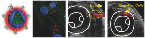
Human embryonic stem cell‐derived cardiomyocytes (hESC‐CMs) have become promising tools to repair injured hearts. To achieve optimal outcomes, advanced molecular imaging methods are essential to accurately track these transplanted cells in the heart. In this study, it is demonstrated for the first time that a class of photoacoustic nanoparticles (PANPs) incorporating semiconducting polymers (SPs) as contrast agents can be used in the photoacoustic imaging (PAI) of transplanted hESC‐CMs in living mouse hearts. This is achieved by virtue of two benefits of PANPs. First, strong photoacoustic (PA) signals and specific spectral features of SPs allow PAI to sensitively detect and distinguish a small number of PANP‐labeled cells (2000) from background tissues. Second, the PANPs show a high efficiency for hESC‐CM labeling without adverse effects on cell structure, function, and gene expression. Assisted by ultrasound imaging, the delivery and engraftment of hESC‐CMs in living mouse hearts can be assessed by PANP‐based PAI with high spatial resolution (≈100 µm). In summary, this study explores and validates a novel application of SPs as a PA contrast agent to track labeled cells with high sensitivity and accuracy in vivo, highlighting the advantages of integrating PAI and PANPs to advance cardiac regenerative therapies.
中文翻译:

使用超灵敏半导体聚合物纳米粒子对活体心脏中胚胎干细胞衍生的心肌细胞进行光声成像
人胚胎干细胞衍生的心肌细胞(hESC-CM)已成为修复受损心脏的有前途的工具。为了实现最佳结果,先进的分子成像方法对于准确追踪心脏中的这些移植细胞至关重要。在这项研究中,首次证明一类包含半导体聚合物(SP)作为造影剂的光声纳米粒子(PANP)可用于活体小鼠心脏中移植的 hESC-CM 的光声成像(PAI)。这是通过 PANP 的两个优点实现的。首先,强光声 (PA) 信号和 SP 的特定光谱特征使 PAI 能够从背景组织中灵敏地检测和区分少量 PANP 标记的细胞 (2000)。其次,PANP 对 hESC-CM 标记显示出高效率,且不会对细胞结构、功能和基因表达产生不利影响。在超声成像的辅助下,hESC-CM 在活体小鼠心脏中的递送和植入可以通过基于 PANP 的 PAI 进行高空间分辨率(约 100 µm)的评估。总之,本研究探索并验证了 SP 作为 PA 造影剂的新应用,可在体内以高灵敏度和准确性追踪标记细胞,突出了整合 PAI 和 PANP 来推进心脏再生治疗的优势。
更新日期:2017-11-08
中文翻译:
使用超灵敏半导体聚合物纳米粒子对活体心脏中胚胎干细胞衍生的心肌细胞进行光声成像
人胚胎干细胞衍生的心肌细胞(hESC-CM)已成为修复受损心脏的有前途的工具。为了实现最佳结果,先进的分子成像方法对于准确追踪心脏中的这些移植细胞至关重要。在这项研究中,首次证明一类包含半导体聚合物(SP)作为造影剂的光声纳米粒子(PANP)可用于活体小鼠心脏中移植的 hESC-CM 的光声成像(PAI)。这是通过 PANP 的两个优点实现的。首先,强光声 (PA) 信号和 SP 的特定光谱特征使 PAI 能够从背景组织中灵敏地检测和区分少量 PANP 标记的细胞 (2000)。其次,PANP 对 hESC-CM 标记显示出高效率,且不会对细胞结构、功能和基因表达产生不利影响。在超声成像的辅助下,hESC-CM 在活体小鼠心脏中的递送和植入可以通过基于 PANP 的 PAI 进行高空间分辨率(约 100 µm)的评估。总之,本研究探索并验证了 SP 作为 PA 造影剂的新应用,可在体内以高灵敏度和准确性追踪标记细胞,突出了整合 PAI 和 PANP 来推进心脏再生治疗的优势。


 京公网安备 11010802027423号