当前位置:
X-MOL 学术
›
Biomater. Sci.
›
论文详情
Our official English website, www.x-mol.net, welcomes your feedback! (Note: you will need to create a separate account there.)
Modified acellular nerve-delivering PMSCs improve functional recovery in rats after complete spinal cord transection
Biomaterials Science ( IF 6.6 ) Pub Date : 2017-11-06 00:00:00 , DOI: 10.1039/c7bm00485k Ting Tian 1, 2, 3, 4 , Zhenhai Yu 1, 2, 3, 4 , Naili Zhang 1, 2, 3, 4 , Yingwei Chang 1, 2, 3, 4 , Yuqiang Zhang 4, 5, 6 , Luping Zhang 1, 2, 3, 4 , Shuai Zhou 1, 2, 3, 4 , Chunlei Zhang 1, 2, 3, 4 , Guoying Feng 1, 2, 3, 4 , Fei Huang 1, 2, 3, 4
Biomaterials Science ( IF 6.6 ) Pub Date : 2017-11-06 00:00:00 , DOI: 10.1039/c7bm00485k Ting Tian 1, 2, 3, 4 , Zhenhai Yu 1, 2, 3, 4 , Naili Zhang 1, 2, 3, 4 , Yingwei Chang 1, 2, 3, 4 , Yuqiang Zhang 4, 5, 6 , Luping Zhang 1, 2, 3, 4 , Shuai Zhou 1, 2, 3, 4 , Chunlei Zhang 1, 2, 3, 4 , Guoying Feng 1, 2, 3, 4 , Fei Huang 1, 2, 3, 4
Affiliation

|
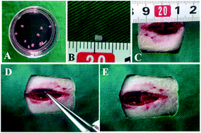
Due to the poor regeneration capacity of neurons and the inhibitory microenvironment, spontaneous regeneration in spinal cord injury (SCI) remains challenging. Tissue engineering is considered a promising approach for enhancing the regeneration of SCI by reconstructing the inherent structure and improving the microenvironment. In this study, the possibility of engineering a nerve complex, which is constructed by acellular nerve delivering placenta mesenchymal stem cells (PMSCs), was assessed for the recovery of a transected spinal cord. Modified acellular nerve grafts were developed, and PMSCs labeled with green fluorescent protein (GFP) were seeded on the graft to construct the engineered nerve complex. Then, the engineered nerve complex was implanted into a 2 mm-length transected gap of the spinal cord. Four weeks after the transplantation, numerous surviving PMSCs were observed in the lesion cavity by immunofluorescence staining. Moreover, co-localization between GFP and neurofilament-200 (NF200) and Neuronal Class III β-Tubulin (Tuj1) was observed at the bridge interface. The PMSCs-graft group exhibited significant function improvement as evaluated by the Basso, Beattie and Bresnahan (BBB) locomotion score and footprint analysis. Eight weeks after surgery, the evoked response was restored in the PMSCs-graft group and numerous thick myelin sheathes were observed compared to that in the control groups. Collectively, our findings suggest that the nerve complex prepared by acellular nerve delivering PMSCs enhanced the structure and function regeneration of the spinal cord after SCI.
中文翻译:

改良的脱细胞神经传递PMSCs改善脊髓完全横断后大鼠的功能恢复
由于神经元的再生能力差和抑制性微环境,脊髓损伤(SCI)的自发再生仍然具有挑战性。组织工程被认为是通过重建固有结构和改善微环境来增强SCI再生的一种有前途的方法。在这项研究中,评估了工程化神经复合物的可能性,该复合物是由无细胞神经传递胎盘间充质干细胞(PMSC)构建的,可用于横断脊髓的恢复。开发了改良的脱细胞神经移植物,并将标记有绿色荧光蛋白(GFP)的PMSC接种在移植物上以构建工程化的神经复合体。然后,将工程神经复合体植入到脊髓的2 mm长横切间隙中。移植四个星期后,通过免疫荧光染色在病变腔中观察到许多存活的PMSC。此外,在桥界面处观察到GFP与神经丝200(NF200)和III类神经元β-微管蛋白(Tuj1)之间的共定位。通过Basso,Beattie和Bresnahan(BBB)的运动评分和足迹分析评估,PMSCs移植组显示出显着的功能改善。术后八周,PMSCs移植组的诱发反应得以恢复,与对照组相比,观察到许多厚的髓鞘。总的来说,我们的发现表明,由脱细胞神经传递的PMSCs制备的神经复合物增强了脊髓损伤后脊髓的结构和功能再生。在桥界面处观察到GFP与神经丝200(NF200)和III类神经元β-微管蛋白(Tuj1)之间的共定位。通过Basso,Beattie和Bresnahan(BBB)的运动评分和足迹分析评估,PMSCs移植组显示出显着的功能改善。术后八周,PMSCs移植组的诱发反应得以恢复,与对照组相比,观察到许多厚的髓鞘。总的来说,我们的发现表明,由脱细胞神经传递的PMSCs制备的神经复合物增强了脊髓损伤后脊髓的结构和功能再生。在桥界面处观察到GFP与神经丝200(NF200)和III类神经元β-微管蛋白(Tuj1)之间的共定位。通过Basso,Beattie和Bresnahan(BBB)的运动评分和足迹分析评估,PMSCs移植组显示出显着的功能改善。术后八周,PMSCs移植组的诱发反应得以恢复,与对照组相比,观察到许多厚的髓鞘。总的来说,我们的发现表明,由脱细胞神经传递的PMSCs制备的神经复合物增强了脊髓损伤后脊髓的结构和功能再生。通过Basso,Beattie和Bresnahan(BBB)的运动评分和足迹分析评估,PMSCs移植组显示出显着的功能改善。术后八周,PMSCs移植组的诱发反应得以恢复,与对照组相比,观察到许多厚的髓鞘。总的来说,我们的发现表明,由脱细胞神经传递的PMSCs制备的神经复合物增强了脊髓损伤后脊髓的结构和功能再生。通过Basso,Beattie和Bresnahan(BBB)的运动评分和足迹分析评估,PMSCs移植组显示出显着的功能改善。术后八周,PMSCs移植组的诱发反应得以恢复,与对照组相比,观察到许多厚的髓鞘。总的来说,我们的发现表明,由脱细胞神经传递的PMSCs制备的神经复合物增强了脊髓损伤后脊髓的结构和功能再生。
更新日期:2017-11-06
中文翻译:

改良的脱细胞神经传递PMSCs改善脊髓完全横断后大鼠的功能恢复
由于神经元的再生能力差和抑制性微环境,脊髓损伤(SCI)的自发再生仍然具有挑战性。组织工程被认为是通过重建固有结构和改善微环境来增强SCI再生的一种有前途的方法。在这项研究中,评估了工程化神经复合物的可能性,该复合物是由无细胞神经传递胎盘间充质干细胞(PMSC)构建的,可用于横断脊髓的恢复。开发了改良的脱细胞神经移植物,并将标记有绿色荧光蛋白(GFP)的PMSC接种在移植物上以构建工程化的神经复合体。然后,将工程神经复合体植入到脊髓的2 mm长横切间隙中。移植四个星期后,通过免疫荧光染色在病变腔中观察到许多存活的PMSC。此外,在桥界面处观察到GFP与神经丝200(NF200)和III类神经元β-微管蛋白(Tuj1)之间的共定位。通过Basso,Beattie和Bresnahan(BBB)的运动评分和足迹分析评估,PMSCs移植组显示出显着的功能改善。术后八周,PMSCs移植组的诱发反应得以恢复,与对照组相比,观察到许多厚的髓鞘。总的来说,我们的发现表明,由脱细胞神经传递的PMSCs制备的神经复合物增强了脊髓损伤后脊髓的结构和功能再生。在桥界面处观察到GFP与神经丝200(NF200)和III类神经元β-微管蛋白(Tuj1)之间的共定位。通过Basso,Beattie和Bresnahan(BBB)的运动评分和足迹分析评估,PMSCs移植组显示出显着的功能改善。术后八周,PMSCs移植组的诱发反应得以恢复,与对照组相比,观察到许多厚的髓鞘。总的来说,我们的发现表明,由脱细胞神经传递的PMSCs制备的神经复合物增强了脊髓损伤后脊髓的结构和功能再生。在桥界面处观察到GFP与神经丝200(NF200)和III类神经元β-微管蛋白(Tuj1)之间的共定位。通过Basso,Beattie和Bresnahan(BBB)的运动评分和足迹分析评估,PMSCs移植组显示出显着的功能改善。术后八周,PMSCs移植组的诱发反应得以恢复,与对照组相比,观察到许多厚的髓鞘。总的来说,我们的发现表明,由脱细胞神经传递的PMSCs制备的神经复合物增强了脊髓损伤后脊髓的结构和功能再生。通过Basso,Beattie和Bresnahan(BBB)的运动评分和足迹分析评估,PMSCs移植组显示出显着的功能改善。术后八周,PMSCs移植组的诱发反应得以恢复,与对照组相比,观察到许多厚的髓鞘。总的来说,我们的发现表明,由脱细胞神经传递的PMSCs制备的神经复合物增强了脊髓损伤后脊髓的结构和功能再生。通过Basso,Beattie和Bresnahan(BBB)的运动评分和足迹分析评估,PMSCs移植组显示出显着的功能改善。术后八周,PMSCs移植组的诱发反应得以恢复,与对照组相比,观察到许多厚的髓鞘。总的来说,我们的发现表明,由脱细胞神经传递的PMSCs制备的神经复合物增强了脊髓损伤后脊髓的结构和功能再生。


 京公网安备 11010802027423号