Journal of Controlled Release ( IF 10.8 ) Pub Date : 2017-10-31 , DOI: 10.1016/j.jconrel.2017.10.042 Katrin Fuchs , Andras Kiss , Pierre E. Bize , Rafael Duran , Alban Denys , Gérard Hopfgartner , Gerrit Borchard , Olivier Jordan
|
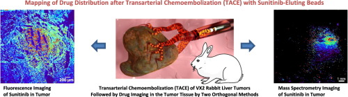
This study describes the use of fluorescence imaging and mass spectrometry imaging, for imaging the anti-angiogenic drug sunitinib, used to treat liver cancer. These techniques allowed for the assessment of local delivery of the unlabeled therapeutic drug. More specifically, the spatial distribution of the drug and its metabolites after local administration was investigated, and drug levels in tumor and liver tissue over time were quantified.
For this purpose, sunitinib-eluting microspheres were locoregionally injected into the tumor feeding arteries of rabbits bearing liver tumors. In adjacent areas of tumor and non-targeted contralateral liver tissue, sunitinib distribution was mapped around beads in occluded vessels 7, 12, 13 and 14 days after embolization by means of the two imaging methods. Presence of sunitinib metabolites was assessed by mass spectrometry imaging.
Sunitinib was found around microspheres in the tumor at day 7, 12, and 13. The drug was retained by the necrotic tumor tissue, resulting in homogeneously distributed and high levels of up to 40 μg/g tissue in a 1.5 mm radius around the beads. The drug was almost completely eliminated from the contralateral liver tissue. Several of the drug's metabolites, including its primary active metabolite SU12662, were detected in the tumor tissue over 13 days.
Sunitinib diffused from the beads and was retained at high, therapeutic levels during 13 days. This was confirmed independently by complementary fluorescence and mass spectrometry imaging, which served as tools to confirm effective drug delivery after hepatic transarterial administration in situ.
Compound: Sunitinib: PubChem CID 5329102.
中文翻译:
互补荧光和质谱成像在兔肝肿瘤模型中的药物分布图
这项研究描述了使用荧光成像和质谱成像技术对用于治疗肝癌的抗血管生成药物舒尼替尼进行成像。这些技术允许评估未标记治疗药物的局部递送。更具体地说,研究了局部给药后药物及其代谢产物的空间分布,并对随着时间推移肿瘤和肝脏组织中的药物水平进行了定量。
为此,将舒尼替尼洗脱的微球局部注射到荷有肝肿瘤的兔子的肿瘤喂养动脉中。在栓塞后第7、12、13和14天,通过两种成像方法,舒尼替尼分布在肿瘤和未靶向的对侧肝组织的相邻区域中,围绕舒尼替尼分布。通过质谱成像评估舒尼替尼代谢物的存在。
在第7、12和13天时,在肿瘤微球周围发现了舒尼替尼。药物被坏死的肿瘤组织保留,导致在珠子周围1.5 mm半径内均匀分布且高水平的高达40μg/ g组织。该药物几乎完全从对侧肝组织中清除。在13天的时间内,在肿瘤组织中检测到该药物的几种代谢物,包括其主要的活性代谢物SU12662。
舒尼替尼从珠子中扩散出来,并在13天内保持较高的治疗水平。补充荧光和质谱成像可独立确认这一点,这是在肝经动脉原位给药后确认有效药物递送的工具。
化合物:舒尼替尼:PubChem CID 5329102。


 京公网安备 11010802027423号