PLoS Pathogens ( IF 6.7 ) Pub Date : 2017-10-17 , DOI: 10.1371/journal.ppat.1006676 Macarena Beigier-Bompadre , Georgina N. Montagna , Anja A. Kühl , Laura Lozza , January Weiner , Andreas Kupz , Alexis Vogelzang , Hans-Joachim Mollenkopf , Delia Löwe , Silke Bandermann , Anca Dorhoi , Volker Brinkmann , Kai Matuschewski , Stefan H. E. Kaufmann
|
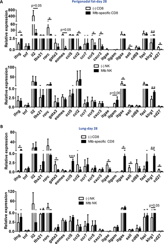
Mycobacterium tuberculosis (Mtb) primarily resides in the lung but can also persist in extrapulmonary sites. Macrophages are considered the prime cellular habitat in all tissues. Here we demonstrate that Mtb resides inside adipocytes of fat tissue where it expresses stress-related genes. Moreover, perigonadal fat of Mtb-infected mice disseminated the infection when transferred to uninfected animals. Adipose tissue harbors leukocytes in addition to adipocytes and other cell types and we observed that Mtb infection induces changes in adipose tissue biology depending on stage of infection. Mice infected via aerosol showed infiltration of inducible nitric oxide synthase (iNOS) or arginase 1 (Arg1)-negative F4/80+ cells, despite recruitment of CD3+, CD4+ and CD8+ T cells. Gene expression analysis of adipose tissue of aerosol Mtb-infected mice provided evidence for upregulated expression of genes associated with T cells and NK cells at 28 days post-infection. Strikingly, IFN-γ-producing NK cells and Mtb-specific CD8+ T cells were identified in perigonadal fat, specifically CD8+CD44-CD69+ and CD8+CD44-CD103+ subpopulations. Gene expression analysis of these cells revealed that they expressed IFN-γ and the lectin-like receptor Klrg1 and down-regulated CD27 and CD62L, consistent with an effector phenotype of Mtb-specific CD8+ T cells. Sorted NK cells expressed higher abundance of Klrg1 upon infection, as well. Our results reveal the ability of Mtb to persist in adipose tissue in a stressed state, and that NK cells and Mtb-specific CD8+ T cells infiltrate infected adipose tissue where they produce IFN-γ and assume an effector phenotype. We conclude that adipose tissue is a potential niche for Mtb and that due to infection CD8+ T cells and NK cells are attracted to this tissue.
中文翻译:
结核分枝杆菌感染调节脂肪组织生物学
结核分枝杆菌(Mtb)主要位于肺中,但也可以在肺外部位持续存在。巨噬细胞被认为是所有组织中主要的细胞栖息地。在这里,我们证明Mtb驻留在脂肪组织的脂肪细胞中,在脂肪细胞中它表达与压力相关的基因。此外,当感染Mtb的小鼠被转移到未感染的动物时,其性腺周围的脂肪传播了感染。除脂肪细胞和其他细胞类型外,脂肪组织还带有白细胞,我们观察到Mtb感染会根据感染的阶段而引起脂肪组织生物学的变化。小鼠通过诱导型一氧化氮合酶的气雾剂显示浸润感染(iNOS)的或精氨酸酶1(ARG1)阴性F4 / 80 +细胞,CD3尽管招募+,CD4 +和CD8 + T细胞。Mtb气溶胶感染小鼠脂肪组织的基因表达分析为感染后28天与T细胞和NK细胞相关的基因表达上调提供了证据。令人惊讶的是,在性腺周围脂肪中,特别是在CD8 + CD44 - CD69 +和CD8 + CD44 - CD103 +亚群中,鉴定出产生IFN-γ的NK细胞和Mtb特异性CD8 + T细胞。这些细胞的基因表达分析表明,它们表达IFN-γ和凝集素样受体Klrg1并下调CD27和CD62L,与Mtb特异性CD8 + T细胞的效应子表型一致。分选的NK细胞在感染时也表达较高的Klrg1丰度。我们的结果揭示了Mtb在压力状态下在脂肪组织中持续存在的能力,并且NK细胞和Mtb特异性CD8 + T细胞浸润感染的脂肪组织,在这些脂肪组织中它们会产生IFN-γ并呈现效应表型。我们得出结论,脂肪组织是Mtb的潜在利基,并且由于感染CD8 + T细胞和NK细胞被吸引到该组织。


 京公网安备 11010802027423号