当前位置:
X-MOL 学术
›
WIREs Comput. Mol. Sci.
›
论文详情
Our official English website, www.x-mol.net, welcomes your feedback! (Note: you will need to create a separate account there.)
3D modeling of chromatin structure: is there a way to integrate and reconcile single cell and population experimental data?
Wiley Interdisciplinary Reviews: Computational Molecular Science ( IF 11.4 ) Pub Date : 2017-04-10 , DOI: 10.1002/wcms.1308 François Le Dily 1, 2 , François Serra 1, 2, 3 , Marc A. Marti-Renom 1, 2, 3, 4
Wiley Interdisciplinary Reviews: Computational Molecular Science ( IF 11.4 ) Pub Date : 2017-04-10 , DOI: 10.1002/wcms.1308 François Le Dily 1, 2 , François Serra 1, 2, 3 , Marc A. Marti-Renom 1, 2, 3, 4
Affiliation

|
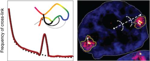
The genome is organized in a hierarchical fashion within the nucleus in interphase. This nonrandom folding of the chromatin fiber is thought to play important roles in the processing of the genetic information. Therefore, a better knowledge of the mechanisms underlying the three‐dimensional structure of the genome appears essential to fully understand the nuclear processes including transcription and replication. Fluorescent in situ hybridization (FISH) and molecular biology methods deriving from the Chromosome Conformation Capture technique are the methods of choice to study genome 3D organization at different levels. Although these single cell and population methods allowed to highlight similar chromatin structures, they also show frequent discrepancies which might be better understood by improving the capacity to generate actual 3D models of organization based on the different types of data available. This review aims at giving an overview of the principles, advantages, and limits of microscopy and molecular biology methods of analysis of genome structure and at discussing the different approaches of modeling of chromatin classically used and the improvements that are necessary to reach a better understanding on the links between chromatin structure and its spatial organization. WIREs Comput Mol Sci 2017, 7:e1308. doi: 10.1002/wcms.1308
中文翻译:

染色质结构的3D建模:是否有一种方法可以整合和协调单细胞和群体实验数据?
基因组在相间的核内以分层的方式组织。染色质纤维的这种非随机折叠被认为在遗传信息的处理中起重要作用。因此,更好地了解基因组三维结构的潜在机制似乎对于充分理解包括转录和复制在内的核过程至关重要。荧光原位染色体构象捕获技术衍生的杂交(FISH)和分子生物学方法是研究不同水平的基因组3D组织的选择方法。尽管这些单细胞和群体方法可以突出显示相似的染色质结构,但它们也显示出频繁的差异,通过提高基于可用数据类型生成实际3D组织模型的能力,可以更好地理解这些差异。这篇综述旨在概述其原理,优势,WIRES Comput Mol Sci 2017,7:e1308。doi:10.1002 / wcms.1308
更新日期:2017-04-10
中文翻译:

染色质结构的3D建模:是否有一种方法可以整合和协调单细胞和群体实验数据?
基因组在相间的核内以分层的方式组织。染色质纤维的这种非随机折叠被认为在遗传信息的处理中起重要作用。因此,更好地了解基因组三维结构的潜在机制似乎对于充分理解包括转录和复制在内的核过程至关重要。荧光原位染色体构象捕获技术衍生的杂交(FISH)和分子生物学方法是研究不同水平的基因组3D组织的选择方法。尽管这些单细胞和群体方法可以突出显示相似的染色质结构,但它们也显示出频繁的差异,通过提高基于可用数据类型生成实际3D组织模型的能力,可以更好地理解这些差异。这篇综述旨在概述其原理,优势,WIRES Comput Mol Sci 2017,7:e1308。doi:10.1002 / wcms.1308


 京公网安备 11010802027423号