Our official English website, www.x-mol.net, welcomes your feedback! (Note: you will need to create a separate account there.)
Plasmonic nanocone arrays for rapid and detailed cell lysate surface enhanced Raman spectroscopy analysis
Analyst ( IF 4.2 ) Pub Date : 2017-10-17 00:00:00 , DOI: 10.1039/c7an00630f L. P. Hackett 1, 2, 3, 4, 5 , L. L. Goddard 1, 2, 3, 4, 5 , G. L. Liu 1, 2, 3, 4, 5
Analyst ( IF 4.2 ) Pub Date : 2017-10-17 00:00:00 , DOI: 10.1039/c7an00630f L. P. Hackett 1, 2, 3, 4, 5 , L. L. Goddard 1, 2, 3, 4, 5 , G. L. Liu 1, 2, 3, 4, 5
Affiliation
|
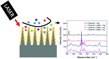
In this work, we develop, fabricate, and characterize a plasmonic nanocone array surface enhanced Raman spectroscopy (SERS) substrate with a uniform enhancement factor on the micron scale for qualitative and quantiative cell and cell lysate analysis. This work demonstrates how SERS substrates can be used as cell-based biosensors given that the enhancement factor of the substrate is sufficient for Raman detection and that the uniformity is high over the applicable surface area. These requirements allow accurate and quantitative comparisons between nonuniform samples under varying biochemical conditions. We apply the developed SERS substrate for Raman measurements and mapping of HeLa cells and cell lysate. This method is used for identification of UV-induced damage and detection of nanomolar concentrations of methylated guanine spiked in cell lysate samples.
中文翻译:

等离子纳米锥阵列,用于快速详细的细胞裂解液表面增强拉曼光谱分析
在这项工作中,我们开发,制造和表征等离子纳米锥阵列表面增强拉曼光谱(SERS)底物,并在微米尺度上具有均一的增强因子,用于定性和定量细胞和细胞裂解物分析。这项工作演示了SERS底物如何用作基于细胞的生物传感器,因为底物的增强因子足以进行拉曼检测,并且在适用表面积上均一性很高。这些要求允许在变化的生化条件下对不均匀样品之间进行准确和定量的比较。我们将开发的SERS底物用于拉曼测量以及HeLa细胞和细胞裂解液的作图。
更新日期:2017-11-20
中文翻译:
等离子纳米锥阵列,用于快速详细的细胞裂解液表面增强拉曼光谱分析
在这项工作中,我们开发,制造和表征等离子纳米锥阵列表面增强拉曼光谱(SERS)底物,并在微米尺度上具有均一的增强因子,用于定性和定量细胞和细胞裂解物分析。这项工作演示了SERS底物如何用作基于细胞的生物传感器,因为底物的增强因子足以进行拉曼检测,并且在适用表面积上均一性很高。这些要求允许在变化的生化条件下对不均匀样品之间进行准确和定量的比较。我们将开发的SERS底物用于拉曼测量以及HeLa细胞和细胞裂解液的作图。


 京公网安备 11010802027423号