当前位置:
X-MOL 学术
›
J. Am. Chem. Soc.
›
论文详情
Our official English website, www.x-mol.net, welcomes your feedback! (Note: you will need to create a separate account there.)
Directly Observing Micelle Fusion and Growth in Solution by Liquid-Cell Transmission Electron Microscopy
Journal of the American Chemical Society ( IF 15.0 ) Pub Date : 2017-11-17 , DOI: 10.1021/jacs.7b09060 Lucas R. Parent 1 , Evangelos Bakalis 2 , Abelardo Ramírez-Hernández 3, 4 , Jacquelin K. Kammeyer 1 , Chiwoo Park 5 , Juan de Pablo 3, 4 , Francesco Zerbetto 2 , Joseph P. Patterson 1, 6 , Nathan C. Gianneschi 1
Journal of the American Chemical Society ( IF 15.0 ) Pub Date : 2017-11-17 , DOI: 10.1021/jacs.7b09060 Lucas R. Parent 1 , Evangelos Bakalis 2 , Abelardo Ramírez-Hernández 3, 4 , Jacquelin K. Kammeyer 1 , Chiwoo Park 5 , Juan de Pablo 3, 4 , Francesco Zerbetto 2 , Joseph P. Patterson 1, 6 , Nathan C. Gianneschi 1
Affiliation

|
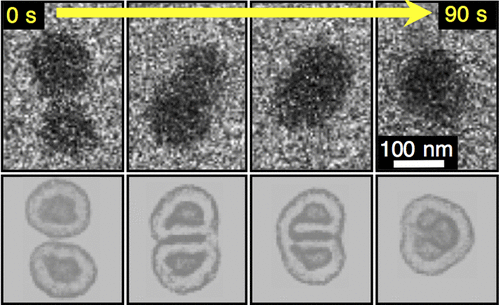
Amphiphilic small molecules and polymers form commonplace nanoscale macromolecular compartments and bilayers, and as such are truly essential components in all cells and in many cellular processes. The nature of these architectures, including their formation, phase changes, and stimuli-response behaviors, is necessary for the most basic functions of life, and over the past half-century, these natural micellar structures have inspired a vast diversity of industrial products, from biomedicines to detergents, lubricants, and coatings. The importance of these materials and their ubiquity have made them the subject of intense investigation regarding their nanoscale dynamics with increasing interest in obtaining sufficient temporal and spatial resolution to directly observe nanoscale processes. However, the vast majority of experimental methods involve either bulk-averaging techniques including light, neutron, and X-ray scattering, or are static in nature including even the most advanced cryogenic transmission electron microscopy techniques. Here, we employ in situ liquid-cell transmission electron microscopy (LCTEM) to directly observe the evolution of individual amphiphilic block copolymer micellar nanoparticles in solution, in real time with nanometer spatial resolution. These observations, made on a proof-of-concept bioconjugate polymer amphiphile, revealed growth and evolution occurring by unimer addition processes and by particle-particle collision-and-fusion events. The experimental approach, combining direct LCTEM observation, quantitative analysis of LCTEM data, and correlated in silico simulations, provides a unique view of solvated soft matter nanoassemblies as they morph and evolve in time and space, enabling us to capture these phenomena in solution.
中文翻译:

通过液体细胞透射电子显微镜直接观察溶液中胶束的融合和生长
两亲性小分子和聚合物形成常见的纳米级大分子隔室和双层,因此是所有细胞和许多细胞过程中真正必不可少的成分。这些结构的性质,包括它们的形成、相变和刺激反应行为,是生命最基本的功能所必需的,在过去的半个世纪里,这些天然胶束结构激发了大量多样化的工业产品,从生物医药到清洁剂、润滑剂和涂料。这些材料的重要性及其普遍性使它们成为对其纳米级动力学进行深入研究的主题,并且越来越有兴趣获得足够的时间和空间分辨率以直接观察纳米级过程。然而,绝大多数实验方法要么涉及体平均技术,包括光、中子和 X 射线散射,要么本质上是静态的,包括最先进的低温透射电子显微镜技术。在这里,我们采用原位液体细胞透射电子显微镜 (LCTEM) 以纳米空间分辨率实时直接观察溶液中单个两亲性嵌段共聚物胶束纳米粒子的演变。这些观察是在概念验证的生物共轭聚合物两亲物上进行的,揭示了由 unimer 添加过程和粒子 - 粒子碰撞和融合事件发生的生长和进化。实验方法,结合直接 LCTEM 观察、LCTEM 数据的定量分析以及相关的计算机模拟,
更新日期:2017-11-17
中文翻译:

通过液体细胞透射电子显微镜直接观察溶液中胶束的融合和生长
两亲性小分子和聚合物形成常见的纳米级大分子隔室和双层,因此是所有细胞和许多细胞过程中真正必不可少的成分。这些结构的性质,包括它们的形成、相变和刺激反应行为,是生命最基本的功能所必需的,在过去的半个世纪里,这些天然胶束结构激发了大量多样化的工业产品,从生物医药到清洁剂、润滑剂和涂料。这些材料的重要性及其普遍性使它们成为对其纳米级动力学进行深入研究的主题,并且越来越有兴趣获得足够的时间和空间分辨率以直接观察纳米级过程。然而,绝大多数实验方法要么涉及体平均技术,包括光、中子和 X 射线散射,要么本质上是静态的,包括最先进的低温透射电子显微镜技术。在这里,我们采用原位液体细胞透射电子显微镜 (LCTEM) 以纳米空间分辨率实时直接观察溶液中单个两亲性嵌段共聚物胶束纳米粒子的演变。这些观察是在概念验证的生物共轭聚合物两亲物上进行的,揭示了由 unimer 添加过程和粒子 - 粒子碰撞和融合事件发生的生长和进化。实验方法,结合直接 LCTEM 观察、LCTEM 数据的定量分析以及相关的计算机模拟,


 京公网安备 11010802027423号