当前位置:
X-MOL 学术
›
Anal. Methods
›
论文详情
Our official English website, www.x-mol.net, welcomes your feedback! (Note: you will need to create a separate account there.)
SmartIHC-Analyzer: smartphone assisted microscopic image analytics for automated Ki-67 quantification in breast cancer evaluation
Analytical Methods ( IF 3.1 ) Pub Date : 2017-10-27 00:00:00 , DOI: 10.1039/c7ay02302b Suman Tewary 1, 2, 3 , Indu Arun 3, 4, 5 , Rosina Ahmed 3, 4, 5 , Sanjoy Chatterjee 3, 4, 5 , Chandan Chakraborty 1, 2, 3
Analytical Methods ( IF 3.1 ) Pub Date : 2017-10-27 00:00:00 , DOI: 10.1039/c7ay02302b Suman Tewary 1, 2, 3 , Indu Arun 3, 4, 5 , Rosina Ahmed 3, 4, 5 , Sanjoy Chatterjee 3, 4, 5 , Chandan Chakraborty 1, 2, 3
Affiliation

|
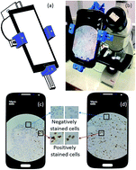
As with other cancers, cell proliferation is one of the indicative hallmarks of breast cancer evaluation. The expression of human Ki-67, being a nuclear protein, has strong association with the proliferation of cancer cells. The proliferation index from Ki-67 is evaluated by immunohistochemical (IHC) study by discriminating positively (brown color) and negatively (blue color) stained nuclei of cells through manual counting. In practice, this evaluation process is highly dependent on an expert-pathologist, as well as being time consuming and prone to inter-observer variability. To circumvent current challenges in IHC image analysis, we introduce SmartIHC-Analyzer, an efficient smartphone assisted microscopy for automatic scoring of Ki-67 protein expression. A universal 3D printed adapter is fabricated to attach a smartphone to the eye-piece of the conventional microscope to acquire microscopic images of stained tissue slides. IHC image acquisition, analytics, visualization and automated reporting for Ki-67 have been developed and integrated in an android smartphone for easy use in any pathological set-up. Color deconvolution followed by morphological top-hat transformation and segmentation is performed to extract stained cells for automated scoring. The proposed SmartIHC-Analyzer app was tested on 30 cases of Ki-67 stained tissue samples and compared with the score given by expert pathologists with variability in illumination conditions. The results had high similarity with manual scoring by the expert pathologists (Pearson's correlation coefficient r = 0.97) and the average absolute variation from pathologists' score was evaluated as 6.21%; which is highly significant. The results have been compared with available state-of-the art ImmunoRatio software where the results are found to be promising. It can be highlighted that SmartIHC-Analyzer can be used in point-of-care diagnostics for instant automatic and augmented reporting of Ki-67 protein expression. The APK file of the SmartIHC-Analyzer is free to use and can be obtained by emailing the corresponding author.
中文翻译:

SmartIHC-Analyzer:智能手机辅助的显微图像分析,用于乳腺癌评估中的自动Ki-67定量
与其他癌症一样,细胞增殖是乳腺癌评估的标志性特征之一。人Ki-67是一种核蛋白,其表达与癌细胞的增殖密切相关。Ki-67的增殖指数通过免疫组化(IHC)研究进行评估,方法是通过人工计数区分阳性(棕色)和阴性(蓝色)染色细胞核。实际上,该评估过程高度依赖于专家病理学家,既耗时又易于观察者之间的差异。为了规避IHC图像分析的当前挑战,我们推出SmartIHC-Analyzer,一种高效的智能手机辅助显微镜,可自动对Ki-67蛋白表达进行评分。制造了通用3D打印适配器,以将智能手机连接到常规显微镜的目镜上,以获取染色的组织玻片的显微图像。针对Ki-67的IHC图像采集,分析,可视化和自动报告已开发并集成在Android智能手机中,可轻松用于任何病理设置中。进行颜色反卷积,然后进行形态学礼帽转换和分割,以提取染色的细胞以进行自动评分。拟议的SmartIHC分析仪在30例Ki-67染色的组织样本上测试了app,并将其与专家病理学家根据照明条件的变化得出的分数进行了比较。结果与专家病理学家的手动评分高度相似(Pearson相关系数r = 0.97),病理学家评分的平均绝对偏差为6.21%;这是非常重要的。将结果与可用的最新ImmunoRatio软件进行了比较,发现结果很有希望。可以强调的是,SmartIHC-Analyzer可用于即时诊断中,以即时自动和增强地报告Ki-67蛋白表达。SmartIHC-Analyzer的APK文件 免费使用,可以通过向相应的作者发送电子邮件获得。
更新日期:2017-11-09
中文翻译:

SmartIHC-Analyzer:智能手机辅助的显微图像分析,用于乳腺癌评估中的自动Ki-67定量
与其他癌症一样,细胞增殖是乳腺癌评估的标志性特征之一。人Ki-67是一种核蛋白,其表达与癌细胞的增殖密切相关。Ki-67的增殖指数通过免疫组化(IHC)研究进行评估,方法是通过人工计数区分阳性(棕色)和阴性(蓝色)染色细胞核。实际上,该评估过程高度依赖于专家病理学家,既耗时又易于观察者之间的差异。为了规避IHC图像分析的当前挑战,我们推出SmartIHC-Analyzer,一种高效的智能手机辅助显微镜,可自动对Ki-67蛋白表达进行评分。制造了通用3D打印适配器,以将智能手机连接到常规显微镜的目镜上,以获取染色的组织玻片的显微图像。针对Ki-67的IHC图像采集,分析,可视化和自动报告已开发并集成在Android智能手机中,可轻松用于任何病理设置中。进行颜色反卷积,然后进行形态学礼帽转换和分割,以提取染色的细胞以进行自动评分。拟议的SmartIHC分析仪在30例Ki-67染色的组织样本上测试了app,并将其与专家病理学家根据照明条件的变化得出的分数进行了比较。结果与专家病理学家的手动评分高度相似(Pearson相关系数r = 0.97),病理学家评分的平均绝对偏差为6.21%;这是非常重要的。将结果与可用的最新ImmunoRatio软件进行了比较,发现结果很有希望。可以强调的是,SmartIHC-Analyzer可用于即时诊断中,以即时自动和增强地报告Ki-67蛋白表达。SmartIHC-Analyzer的APK文件 免费使用,可以通过向相应的作者发送电子邮件获得。


 京公网安备 11010802027423号