当前位置:
X-MOL 学术
›
Acta Mater.
›
论文详情
Our official English website, www.x-mol.net, welcomes your feedback! (Note: you will need to create a separate account there.)
Stress-induced microcrack density evolution in β-eucryptite ceramics: experimental observations and possible route to strain hardening
Acta Materialia ( IF 9.4 ) Pub Date : 2018-02-01 , DOI: 10.1016/j.actamat.2017.10.030 B.R. Müller , R.C. Cooper , A. Lange , A. Kupsch , M. Wheeler , M.P. Hentschel , A. Staude , A. Pandey , A. Shyam , G. Bruno
Acta Materialia ( IF 9.4 ) Pub Date : 2018-02-01 , DOI: 10.1016/j.actamat.2017.10.030 B.R. Müller , R.C. Cooper , A. Lange , A. Kupsch , M. Wheeler , M.P. Hentschel , A. Staude , A. Pandey , A. Shyam , G. Bruno
|
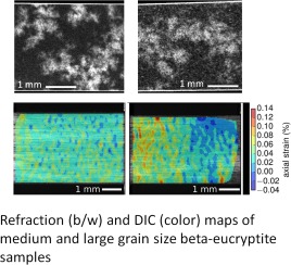
Abstract In order to investigate their microcracking behaviour, the microstructures of several β-eucryptite ceramics, obtained from glass precursor and cerammed to yield different grain sizes and microcrack densities, were characterized by laboratory and synchrotron x-ray refraction and tomography. Results were compared with those obtained from scanning electron microscopy (SEM). In SEM images, the characterized materials appeared fully dense but computed tomography showed the presence of pore clusters. Uniaxial tensile testing was performed on specimens while strain maps were recorded and analyzed by Digital Image Correlation (DIC). X-ray refraction techniques were applied on specimens before and after tensile testing to measure the amount of the internal specific surface (i.e., area per unit volume). X-ray refraction revealed that (a) the small grain size (SGS) material contained a large specific surface, originating from the grain boundaries and the interfaces of TiO2 precipitates; (b) the medium (MGS) and large grain size (LGS) materials possessed higher amounts of specific surface compared to SGS material due to microcracks, which decreased after tensile loading; (c) the precursor glass had negligible internal surface. The unexpected decrease in the internal surface of MGS and LGS after tensile testing is explained by the presence of compressive regions in the DIC strain maps and further by theoretical arguments. It is suggested that while some microcracks merge via propagation, more close mechanically, thereby explaining the observed X-ray refraction results. The mechanisms proposed would allow the development of a strain hardening route in ceramics.
中文翻译:

β-锂霞石陶瓷中应力诱导的微裂纹密度演变:实验观察和应变硬化的可能途径
摘要 为了研究它们的微裂纹行为,从玻璃前驱体中获得并陶瓷化以产生不同晶粒尺寸和微裂纹密度的几种 β-锂霞石陶瓷的微观结构,通过实验室和同步加速器 X 射线折射和断层扫描进行了表征。结果与从扫描电子显微镜 (SEM) 获得的结果进行了比较。在 SEM 图像中,表征材料看起来完全致密,但计算机断层扫描显示存在孔簇。对试样进行单轴拉伸试验,同时通过数字图像相关 (DIC) 记录和分析应变图。在拉伸试验之前和之后,将 X 射线折射技术应用于试样以测量内部比表面积(即,每单位体积的面积)的量。X 射线折射显示 (a) 小晶粒尺寸 (SGS) 材料包含大比表面积,源自晶粒边界和 TiO2 沉淀物的界面;(b) 与 SGS 材料相比,中等 (MGS) 和大晶粒尺寸 (LGS) 材料具有更高的比表面积,因为微裂纹在拉伸加载后会减少;(c) 前体玻璃的内表面可以忽略不计。拉伸测试后 MGS 和 LGS 内表面的意外减少可以通过 DIC 应变图中压缩区域的存在以及理论论证来解释。建议虽然一些微裂纹通过传播合并,但机械上更接近,从而解释了观察到的 X 射线折射结果。
更新日期:2018-02-01
中文翻译:
β-锂霞石陶瓷中应力诱导的微裂纹密度演变:实验观察和应变硬化的可能途径
摘要 为了研究它们的微裂纹行为,从玻璃前驱体中获得并陶瓷化以产生不同晶粒尺寸和微裂纹密度的几种 β-锂霞石陶瓷的微观结构,通过实验室和同步加速器 X 射线折射和断层扫描进行了表征。结果与从扫描电子显微镜 (SEM) 获得的结果进行了比较。在 SEM 图像中,表征材料看起来完全致密,但计算机断层扫描显示存在孔簇。对试样进行单轴拉伸试验,同时通过数字图像相关 (DIC) 记录和分析应变图。在拉伸试验之前和之后,将 X 射线折射技术应用于试样以测量内部比表面积(即,每单位体积的面积)的量。X 射线折射显示 (a) 小晶粒尺寸 (SGS) 材料包含大比表面积,源自晶粒边界和 TiO2 沉淀物的界面;(b) 与 SGS 材料相比,中等 (MGS) 和大晶粒尺寸 (LGS) 材料具有更高的比表面积,因为微裂纹在拉伸加载后会减少;(c) 前体玻璃的内表面可以忽略不计。拉伸测试后 MGS 和 LGS 内表面的意外减少可以通过 DIC 应变图中压缩区域的存在以及理论论证来解释。建议虽然一些微裂纹通过传播合并,但机械上更接近,从而解释了观察到的 X 射线折射结果。


 京公网安备 11010802027423号