当前位置:
X-MOL 学术
›
Acc. Chem. Res.
›
论文详情
Our official English website, www.x-mol.net, welcomes your feedback! (Note: you will need to create a separate account there.)
Application of X-ray Diffraction and Electron Crystallography for Solving Complex Structure Problems
Accounts of Chemical Research ( IF 18.3 ) Pub Date : 2017-11-01 00:00:00 , DOI: 10.1021/acs.accounts.7b00366 Jian Li 1 , Junliang Sun 1
Accounts of Chemical Research ( IF 18.3 ) Pub Date : 2017-11-01 00:00:00 , DOI: 10.1021/acs.accounts.7b00366 Jian Li 1 , Junliang Sun 1
Affiliation
|
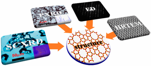
All crystalline materials in nature, whether inorganic, organic, or biological, macroscopic or microscopic, have their own chemical and physical properties, which strongly depend on their atomic structures. Therefore, structure determination is extremely important in chemistry, physics, materials science, etc. In the past centuries, many techniques have been developed for structure determination. The most widely used one is X-ray crystallography (single-crystal X-ray diffraction (SCXRD) and powder X-ray diffraction (PXRD)), and it remains the most important technique for structure determination of crystalline materials. Although SCXRD and PXRD are successful in many cases, a number of reasons limit their applications, such as SCXRD for nanosized crystals, intergrowth, and defects and PXRD for complex structures, multiphasic samples, impurities, peak overlaps, etc. Another most valuable technique for structure determination is electron crystallography (EC). With the electron as a probe, EC alone can also be used for structure determination, especially for crystals that are too small to be studied by SCXRD or too complex for PXRD. As electrons interact much more strongly with matter than X-rays do, both electron diffraction (ED) patterns and high-resolution transmission electron microscopy (HRTEM) images can be obtained from nanosized crystals. However, collecting a complete set of ED patterns or recording a good HRTEM image requires considerable expertise on the operation of electron microscopes and crystallography. The strong interactions between electrons and materials can also lead to dynamical effects and beam damage. These difficulties make structure determination from ED patterns and HRTEM images not straightforward. Recently, two three-dimensional (3D) electron diffraction techniques, automated electron diffraction tomography (ADT) and rotation electron diffraction (RED), have been developed, which perform the data collection in an automated manner. Although the dynamical effects in the newly developed 3D electron diffraction techniques (ADT, RED) are reduced significantly, for some structures there are still problems with obtaining an initial model because of beam damage.
中文翻译:

X射线衍射和电子晶体学在解决复杂结构问题中的应用
自然界中的所有晶体材料,无论是无机的,有机的,生物的,宏观的还是微观的,都具有自己的化学和物理性质,这在很大程度上取决于其原子结构。因此,结构确定在化学,物理学,材料科学等领域极为重要。在过去的几个世纪中,已经开发了许多用于结构确定的技术。使用最广泛的一种是X射线晶体学(单晶X射线衍射(SCXRD)和粉末X射线衍射(PXRD)),它仍然是确定晶体材料结构的最重要技术。尽管SCXRD和PXRD在许多情况下都成功了,但是有许多原因限制了它们的应用,例如SCXRD用于纳米级晶体,共生和缺陷,而PXRD用于复杂结构,多相样品,杂质,峰重叠等。结构确定的另一种最有价值的技术是电子晶体学(EC)。以电子作为探针,单独的EC也可以用于结构确定,特别是对于那些太小而无法通过SCXRD研究的晶体或对于PXRD太复杂的晶体。由于电子与物质的相互作用比X射线强烈得多,因此可以从纳米级晶体获得电子衍射(ED)图和高分辨率透射电子显微镜(HRTEM)图像。但是,收集一套完整的ED模式或记录良好的HRTEM图像需要在电子显微镜和晶体学操作方面有大量的专业知识。电子与材料之间的强相互作用也可能导致动力学效应和电子束损坏。这些困难使得从ED模式和HRTEM图像确定结构变得不容易。最近,已经开发了两种三维(3D)电子衍射技术,即自动电子衍射层析成像(ADT)和旋转电子衍射(RED),它们以自动化方式执行数据收集。尽管新开发的3D电子衍射技术(ADT,RED)的动力学效果已大大降低,但对于某些结构,由于束损坏,仍然存在获取初始模型的问题。
更新日期:2017-11-01
中文翻译:
X射线衍射和电子晶体学在解决复杂结构问题中的应用
自然界中的所有晶体材料,无论是无机的,有机的,生物的,宏观的还是微观的,都具有自己的化学和物理性质,这在很大程度上取决于其原子结构。因此,结构确定在化学,物理学,材料科学等领域极为重要。在过去的几个世纪中,已经开发了许多用于结构确定的技术。使用最广泛的一种是X射线晶体学(单晶X射线衍射(SCXRD)和粉末X射线衍射(PXRD)),它仍然是确定晶体材料结构的最重要技术。尽管SCXRD和PXRD在许多情况下都成功了,但是有许多原因限制了它们的应用,例如SCXRD用于纳米级晶体,共生和缺陷,而PXRD用于复杂结构,多相样品,杂质,峰重叠等。结构确定的另一种最有价值的技术是电子晶体学(EC)。以电子作为探针,单独的EC也可以用于结构确定,特别是对于那些太小而无法通过SCXRD研究的晶体或对于PXRD太复杂的晶体。由于电子与物质的相互作用比X射线强烈得多,因此可以从纳米级晶体获得电子衍射(ED)图和高分辨率透射电子显微镜(HRTEM)图像。但是,收集一套完整的ED模式或记录良好的HRTEM图像需要在电子显微镜和晶体学操作方面有大量的专业知识。电子与材料之间的强相互作用也可能导致动力学效应和电子束损坏。这些困难使得从ED模式和HRTEM图像确定结构变得不容易。最近,已经开发了两种三维(3D)电子衍射技术,即自动电子衍射层析成像(ADT)和旋转电子衍射(RED),它们以自动化方式执行数据收集。尽管新开发的3D电子衍射技术(ADT,RED)的动力学效果已大大降低,但对于某些结构,由于束损坏,仍然存在获取初始模型的问题。


 京公网安备 11010802027423号