当前位置:
X-MOL 学术
›
Chem. Sci.
›
论文详情
Our official English website, www.x-mol.net, welcomes your feedback! (Note: you will need to create a separate account there.)
3D chemical imaging of the brain using quantitative IR spectro-microscopy
Chemical Science ( IF 8.4 ) Pub Date : 2017-10-17 00:00:00 , DOI: 10.1039/c7sc03306k Abiodun Ogunleke 1, 2, 3 , Benoit Recur 1, 2, 3 , Hugo Balacey 1, 2, 3 , Hsiang-Hsin Chen 4, 5, 6, 7 , Maylis Delugin 1, 2, 3 , Yeukuang Hwu 4, 5, 6, 7 , Sophie Javerzat 1, 2, 3 , Cyril Petibois 1, 2, 3, 4, 5
Chemical Science ( IF 8.4 ) Pub Date : 2017-10-17 00:00:00 , DOI: 10.1039/c7sc03306k Abiodun Ogunleke 1, 2, 3 , Benoit Recur 1, 2, 3 , Hugo Balacey 1, 2, 3 , Hsiang-Hsin Chen 4, 5, 6, 7 , Maylis Delugin 1, 2, 3 , Yeukuang Hwu 4, 5, 6, 7 , Sophie Javerzat 1, 2, 3 , Cyril Petibois 1, 2, 3, 4, 5
Affiliation
|
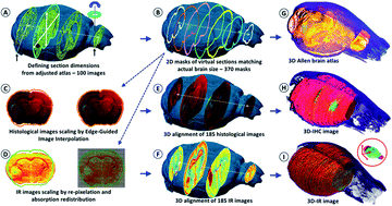
Three-dimensional (3D) histology is the next frontier for modern anatomo-pathology. Characterizing abnormal parameters in a tissue is essential to understand the rationale of pathology development. However, there is no analytical technique, in vivo or histological, that is able to discover such abnormal features and provide a 3D distribution at microscopic resolution. Here, we introduce a unique high-throughput infrared (IR) microscopy method that combines automated image correction and subsequent spectral data analysis for 3D-IR image reconstruction. We performed spectral analysis of a complete organ for a small animal model, a mouse brain with an implanted glioma tumor. The 3D-IR image is reconstructed from 370 consecutive tissue sections and corrected using the X-ray tomogram of the organ for an accurate quantitative analysis of the chemical content. A 3D matrix of 89 × 106 IR spectra is generated, allowing us to separate the tumor mass from healthy brain tissues based on various anatomical, chemical, and metabolic parameters. We demonstrate that quantitative metabolic parameters can be extracted from the IR spectra for the characterization of the brain vs. tumor metabolism (assessing the Warburg effect in tumors). Our method can be further exploited by searching for the whole spectral profile, discriminating tumor vs. healthy tissue in a non-supervised manner, which we call ‘spectromics’.
中文翻译:

使用定量红外光谱显微镜对大脑进行3D化学成像
三维(3D)组织学是现代解剖病理学的下一个前沿领域。表征组织中的异常参数对于理解病理学发展的基本原理至关重要。但是,体内没有分析技术或组织学上能够发现此类异常特征并提供微观分辨率的3D分布。在这里,我们介绍了一种独特的高通量红外(IR)显微镜方法,该方法结合了自动图像校正和后续光谱数据分析的3D-IR图像重建功能。我们对小动物模型(具有植入的神经胶质瘤肿瘤的小鼠大脑)的完整器官进行了光谱分析。从370个连续的组织切片中重建3D-IR图像,并使用器官的X射线断层图进行校正,以对化学成分进行准确的定量分析。89×10 6的3D矩阵生成红外光谱,使我们能够基于各种解剖学,化学和代谢参数将肿瘤块与健康的脑组织分开。我们证明,可以从红外光谱中提取定量代谢参数,以表征大脑与肿瘤代谢的关系(评估肿瘤中的Warburg效应)。通过搜索整个光谱图,以非监督方式(我们称为“光谱学”)区分肿瘤与健康组织,可以进一步利用我们的方法。
更新日期:2017-11-13
中文翻译:
使用定量红外光谱显微镜对大脑进行3D化学成像
三维(3D)组织学是现代解剖病理学的下一个前沿领域。表征组织中的异常参数对于理解病理学发展的基本原理至关重要。但是,体内没有分析技术或组织学上能够发现此类异常特征并提供微观分辨率的3D分布。在这里,我们介绍了一种独特的高通量红外(IR)显微镜方法,该方法结合了自动图像校正和后续光谱数据分析的3D-IR图像重建功能。我们对小动物模型(具有植入的神经胶质瘤肿瘤的小鼠大脑)的完整器官进行了光谱分析。从370个连续的组织切片中重建3D-IR图像,并使用器官的X射线断层图进行校正,以对化学成分进行准确的定量分析。89×10 6的3D矩阵生成红外光谱,使我们能够基于各种解剖学,化学和代谢参数将肿瘤块与健康的脑组织分开。我们证明,可以从红外光谱中提取定量代谢参数,以表征大脑与肿瘤代谢的关系(评估肿瘤中的Warburg效应)。通过搜索整个光谱图,以非监督方式(我们称为“光谱学”)区分肿瘤与健康组织,可以进一步利用我们的方法。


 京公网安备 11010802027423号