当前位置:
X-MOL 学术
›
Mol. Pharmaceutics
›
论文详情
Our official English website, www.x-mol.net, welcomes your feedback! (Note: you will need to create a separate account there.)
Characterization of Pegylated Liposomal Mitomycin C Lipid-Based Prodrug (Promitil) by High Sensitivity Differential Scanning Calorimetry and Cryogenic Transmission Electron Microscopy
Molecular Pharmaceutics ( IF 4.9 ) Pub Date : 2017-10-30 00:00:00 , DOI: 10.1021/acs.molpharmaceut.6b00865 Xiaohui Wei 1, 2 , Yogita Patil 3, 4 , Patricia Ohana 5 , Yasmine Amitay 5 , Hilary Shmeeda 3 , Alberto Gabizon 3, 4, 5 , Yechezkel Barenholz 1
Molecular Pharmaceutics ( IF 4.9 ) Pub Date : 2017-10-30 00:00:00 , DOI: 10.1021/acs.molpharmaceut.6b00865 Xiaohui Wei 1, 2 , Yogita Patil 3, 4 , Patricia Ohana 5 , Yasmine Amitay 5 , Hilary Shmeeda 3 , Alberto Gabizon 3, 4, 5 , Yechezkel Barenholz 1
Affiliation
|
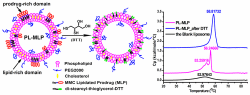
The effect of a lipidated prodrug of mitomycin C (MLP) on the membrane of a pegylated liposome formulation (PL-MLP), also known as Promitil, was characterized through high-sensitivity differential scanning calorimetry (DSC) and cryo-TEM. The thermodynamic analysis demonstrated that MLP led to the formation of heterogeneous domains in the membrane plane of PL-MLP. MLP concentrated in prodrug-rich domains, arranged in high-ordered crystal-like structures, as suggested by the sharp and high enthalpy endotherm in the first heating scanning. After thiolytic cleavage of mitomycin C from MLP by dithiothreitol (DTT) treatment, the crystal-like prodrug domain disappears and a homogeneous membrane with stronger lipid interactions and higher phase transition temperature compared with the blank (MLP-free) liposomes is observed by DSC. In parallel, the rod-like discoid liposomes and the “kissing liposomes” seen by cryo-TEM in the PL-MLP formulation disappear, and liposome mean size and polydispersity increase after DTT treatment. Both MLP and the residual postcleavage lipophilic moiety of the prodrug increased the rigidity of the liposome membrane as indicated by DSC. These results confirm that MLP is inserted in the PL-MLP liposome membrane via its lipophilic anchor, and its mitomycin C moiety located mainly at the region of the phospholipid glycerol backbone and polar headgroup. We hypothesize that π–π stacking between the planar aromatic rings of the mitomycin C moieties leads to the formation of prodrug-rich domains with highly ordered structure on the PL-MLP liposome membrane. This thermodynamically stable conformation may explain the high stability of the PL-MLP formulation. These results also provide us with an interesting example of the application of high sensitivity DSC in understanding the composition–structure–behavior dynamics of liposomal nanocarriers having a lipid-based drug as pharmaceutical ingredient.
中文翻译:

高灵敏度差示扫描量热法和低温透射电镜表征聚乙二醇化脂质体丝裂霉素C脂质基前药(普罗米蒂)
丝裂霉素C(MLP)的脂质化前药对聚乙二醇化脂质体制剂(PL-MLP)(也称为Promitil)膜的作用通过高灵敏度差示扫描量热法(DSC)和冷冻TEM进行了表征。热力学分析表明,MLP导致PL-MLP膜平面中异质域的形成。MLP集中在前药丰富的区域中,排列成高阶晶体状结构,这在第一次加热扫描中就表现出尖锐而高的焓吸热。通过二硫苏糖醇(DTT)处理从MLP进行丝裂霉素C的硫解酶切后,与空白(不含MLP的)脂质体相比,通过DSC观察到晶体状的前药结构域消失,脂质相互作用和相变温度更高的均质膜。在平行下,冷冻TEM在PL-MLP制剂中观察到的棒状盘状脂质体和“亲吻脂质体”消失,DTT处理后脂质体平均大小和多分散性增加。如DSC所示,MLP和前药的残余裂解后亲脂部分均增加了脂质体膜的刚性。这些结果证实了MLP通过其亲脂性锚和其丝裂霉素C部分主要位于磷脂甘油主链和极性头基的区域插入到PL-MLP脂质体膜中。我们假设丝裂霉素C部分的平面芳香环之间存在π-π堆积,从而导致PL-MLP脂质体膜上形成高度有序结构的富含前药的结构域。该热力学稳定的构象可以解释PL-MLP制剂的高稳定性。
更新日期:2017-10-30
中文翻译:
高灵敏度差示扫描量热法和低温透射电镜表征聚乙二醇化脂质体丝裂霉素C脂质基前药(普罗米蒂)
丝裂霉素C(MLP)的脂质化前药对聚乙二醇化脂质体制剂(PL-MLP)(也称为Promitil)膜的作用通过高灵敏度差示扫描量热法(DSC)和冷冻TEM进行了表征。热力学分析表明,MLP导致PL-MLP膜平面中异质域的形成。MLP集中在前药丰富的区域中,排列成高阶晶体状结构,这在第一次加热扫描中就表现出尖锐而高的焓吸热。通过二硫苏糖醇(DTT)处理从MLP进行丝裂霉素C的硫解酶切后,与空白(不含MLP的)脂质体相比,通过DSC观察到晶体状的前药结构域消失,脂质相互作用和相变温度更高的均质膜。在平行下,冷冻TEM在PL-MLP制剂中观察到的棒状盘状脂质体和“亲吻脂质体”消失,DTT处理后脂质体平均大小和多分散性增加。如DSC所示,MLP和前药的残余裂解后亲脂部分均增加了脂质体膜的刚性。这些结果证实了MLP通过其亲脂性锚和其丝裂霉素C部分主要位于磷脂甘油主链和极性头基的区域插入到PL-MLP脂质体膜中。我们假设丝裂霉素C部分的平面芳香环之间存在π-π堆积,从而导致PL-MLP脂质体膜上形成高度有序结构的富含前药的结构域。该热力学稳定的构象可以解释PL-MLP制剂的高稳定性。


 京公网安备 11010802027423号