当前位置:
X-MOL 学术
›
Mater. Horiz.
›
论文详情
Our official English website, www.x-mol.net, welcomes your feedback! (Note: you will need to create a separate account there.)
LiGa5O8:Cr-based theranostic nanoparticles for imaging-guided X-ray induced photodynamic therapy of deep-seated tumors
Materials Horizons ( IF 13.3 ) Pub Date : 2017-08-09 00:00:00 , DOI: 10.1039/c7mh00442g Hongmin Chen 1, 2, 3, 4, 5 , Xilin Sun 1, 2, 3, 4, 5 , Geoffrey D. Wang 6, 7, 8, 9 , Koichi Nagata 7, 8, 9, 10 , Zhonglin Hao 11, 12, 13, 14, 15 , Andrew Wang 9, 16, 17, 18, 19 , Zibo Li 9, 18, 19, 20 , Jin Xie 6, 7, 8, 9 , Baozhong Shen 1, 2, 3, 4, 5
Materials Horizons ( IF 13.3 ) Pub Date : 2017-08-09 00:00:00 , DOI: 10.1039/c7mh00442g Hongmin Chen 1, 2, 3, 4, 5 , Xilin Sun 1, 2, 3, 4, 5 , Geoffrey D. Wang 6, 7, 8, 9 , Koichi Nagata 7, 8, 9, 10 , Zhonglin Hao 11, 12, 13, 14, 15 , Andrew Wang 9, 16, 17, 18, 19 , Zibo Li 9, 18, 19, 20 , Jin Xie 6, 7, 8, 9 , Baozhong Shen 1, 2, 3, 4, 5
Affiliation
|
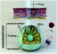
Using X-rays as the irradiation source, a photodynamic therapy process can be initiated in deep tissues. This technology, referred to as X-ray induced PDT, or X-PDT, holds great potential to treat tumors in internal organs. To this end, one question is how to navigate the treatment of tumors with accuracy using external irradiation. Herein we address this issue using a novel LiGa5O8:Cr (LGO:Cr)-based nanoscintillator, which emits persistent, near-infrared X-ray luminescence. This permits deep-tissue optical imaging that can be employed to guide irradiation. Specifically, we encapsulated LGO:Cr nanoparticles and a photosensitizer, 2,3-naphthalocyanine, into mesoporous silica nanoparticles. The nanoparticles were conjugated with cetuximab and systemically injected into H1299 orthotopic non-small cell lung cancer tumor models. The nanoconjugates can efficiently accumulate in tumors in the lungs, confirmed by monitoring the X-ray luminescence from LGO:Cr. Guided by the imaging, external irradiation was applied, leading to efficient tumor suppression while minimally affecting normal tissues. To the best of our knowledge, the present study is the first to demonstrate, with systematically injected nanoparticles, that X-PDT can suppress the growth of deep-seated tumors. The imaging guidance is also new to X-PDT, and is significant to the further transformation of the technology.
中文翻译:

LiGa 5 O 8:Cr基治疗性纳米粒子,用于影像引导X射线诱导的深部肿瘤光动力治疗
使用X射线作为辐照源,可以在深层组织中启动光动力疗法。这项技术被称为X射线诱导的PDT或X-PDT,在治疗内部器官肿瘤方面具有巨大潜力。为此,一个问题是如何使用外部照射来精确地指导肿瘤的治疗。本文中,我们使用新型LiGa 5 O 8解决了这个问题:Cr(LGO:Cr)基纳米闪烁体,它发出持续的近红外X射线发光。这允许可用于引导辐射的深部组织光学成像。具体而言,我们将LGO:Cr纳米颗粒和光敏剂2,3-萘酞菁包封到中孔二氧化硅纳米颗粒中。将纳米颗粒与西妥昔单抗缀合并全身注射到H1299原位非小细胞肺癌肿瘤模型中。通过监测LGO:Cr的X射线发光证实,纳米共轭物可以有效地在肺部肿瘤中蓄积。在成像的指导下,应用外部照射,从而在有效地抑制肿瘤的同时将对正常组织的影响降至最低。据我们所知,本研究是第一个系统注射纳米颗粒的实验,X-PDT可以抑制深层肿瘤的生长。成像指导对于X-PDT来说也是新的,对技术的进一步转变具有重要意义。
更新日期:2017-10-30
中文翻译:
LiGa 5 O 8:Cr基治疗性纳米粒子,用于影像引导X射线诱导的深部肿瘤光动力治疗
使用X射线作为辐照源,可以在深层组织中启动光动力疗法。这项技术被称为X射线诱导的PDT或X-PDT,在治疗内部器官肿瘤方面具有巨大潜力。为此,一个问题是如何使用外部照射来精确地指导肿瘤的治疗。本文中,我们使用新型LiGa 5 O 8解决了这个问题:Cr(LGO:Cr)基纳米闪烁体,它发出持续的近红外X射线发光。这允许可用于引导辐射的深部组织光学成像。具体而言,我们将LGO:Cr纳米颗粒和光敏剂2,3-萘酞菁包封到中孔二氧化硅纳米颗粒中。将纳米颗粒与西妥昔单抗缀合并全身注射到H1299原位非小细胞肺癌肿瘤模型中。通过监测LGO:Cr的X射线发光证实,纳米共轭物可以有效地在肺部肿瘤中蓄积。在成像的指导下,应用外部照射,从而在有效地抑制肿瘤的同时将对正常组织的影响降至最低。据我们所知,本研究是第一个系统注射纳米颗粒的实验,X-PDT可以抑制深层肿瘤的生长。成像指导对于X-PDT来说也是新的,对技术的进一步转变具有重要意义。


 京公网安备 11010802027423号