Our official English website, www.x-mol.net, welcomes your feedback! (Note: you will need to create a separate account there.)
A combined experimental and theoretical approach towards mechanophenotyping of biological cells using a constricted microchannel
Lab on a Chip ( IF 6.1 ) Pub Date : 2017-09-27 00:00:00 , DOI: 10.1039/c7lc00599g A. Raj 1, 2, 3, 4 , M. Dixit 2, 3, 4, 5 , M. Doble 2, 3, 4, 5 , A. K. Sen 1, 2, 3, 4
Lab on a Chip ( IF 6.1 ) Pub Date : 2017-09-27 00:00:00 , DOI: 10.1039/c7lc00599g A. Raj 1, 2, 3, 4 , M. Dixit 2, 3, 4, 5 , M. Doble 2, 3, 4, 5 , A. K. Sen 1, 2, 3, 4
Affiliation
|
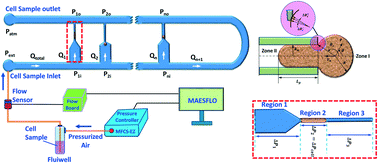
We report a combined experimental and theoretical technique that enables the characterization of various mechanical properties of biological cells. The cells were infused into a microfluidic device that comprises multiple parallel micro-constrictions to eliminate device clogging and facilitate characterization of cells of different sizes and types on a single device. The extension ratio λ and transit velocity Uc of the cells were measured using high-speed and high-resolution imaging which were then used in a theoretical model to predict the Young's modulus Ec = f(λ, Uc) of the cells. The predicted Young's modulus Ec values for three different cell lines (182 ± 34.74 Pa for MDA MB 231, 360 ± 75 Pa for MCF 10A and, 763 ± 93 Pa for HeLa) compare well with those reported in the literature from micropipette measurements and atomic force microscopy measurement within 10% and 15%, respectively. Also, the Young's modulus of MDA-MB-231 cells treated with 50 μM 4-hyrdroxyacetophenone (for localization of myosin II) for 30 min was found out to be 260 ± 52 Pa. The entry time te of cells into the micro-constrictions was predicted using the model and validated using experimentally measured data. The entry and transit behaviors of cells in the micro-constriction including cell deformation (extension ratio λ) and velocity Uc were experimentally measured and used to predict various cell properties such as the Young's modulus, cytoplasmic viscosity and induced hydrodynamic resistance of different types of cells. The proposed combined experimental and theoretical approach leads to a new paradigm for mechanophenotyping of biological cells.
中文翻译:

结合实验和理论方法使用狭窄的微通道对生物细胞进行机械分型
我们报告了结合的实验和理论技术,使生物细胞的各种机械性能的表征。将细胞注入包含多个平行微缩孔的微流控设备中,以消除设备堵塞,并有助于在单个设备上表征不同大小和类型的细胞。使用高速和高分辨率成像测量细胞的延伸率λ和通过速度U c,然后将其用于理论模型中以预测细胞的杨氏模量E c = f(λ,U c)。预测杨氏模量E c三种不同细胞系的值(MDA MB 231为182±34.74 Pa,MCF 10A为360±75 Pa,HeLa为763±93 Pa)与文献报道的微量移液器测量和原子力显微镜测量结果在10 %和15%。此外,MDA-MB-231细胞的杨氏模量用50μM4- hyrdroxyacetophenone(肌球蛋白II的定位)处理30分钟,被发现是260±52帕。该条目时间吨Ë细胞进入微使用模型预测收缩,并使用实验测量的数据进行验证。细胞在微收缩区的进入和迁移行为,包括细胞变形(延伸率λ)和速度U c通过实验测量并用于预测各种细胞特性,例如杨氏模量,细胞质粘度和不同类型细胞的诱导的流体动力学阻力。提出的实验和理论相结合的方法导致了生物细胞机械表型的新范式。
更新日期:2017-10-26
中文翻译:
结合实验和理论方法使用狭窄的微通道对生物细胞进行机械分型
我们报告了结合的实验和理论技术,使生物细胞的各种机械性能的表征。将细胞注入包含多个平行微缩孔的微流控设备中,以消除设备堵塞,并有助于在单个设备上表征不同大小和类型的细胞。使用高速和高分辨率成像测量细胞的延伸率λ和通过速度U c,然后将其用于理论模型中以预测细胞的杨氏模量E c = f(λ,U c)。预测杨氏模量E c三种不同细胞系的值(MDA MB 231为182±34.74 Pa,MCF 10A为360±75 Pa,HeLa为763±93 Pa)与文献报道的微量移液器测量和原子力显微镜测量结果在10 %和15%。此外,MDA-MB-231细胞的杨氏模量用50μM4- hyrdroxyacetophenone(肌球蛋白II的定位)处理30分钟,被发现是260±52帕。该条目时间吨Ë细胞进入微使用模型预测收缩,并使用实验测量的数据进行验证。细胞在微收缩区的进入和迁移行为,包括细胞变形(延伸率λ)和速度U c通过实验测量并用于预测各种细胞特性,例如杨氏模量,细胞质粘度和不同类型细胞的诱导的流体动力学阻力。提出的实验和理论相结合的方法导致了生物细胞机械表型的新范式。


 京公网安备 11010802027423号