当前位置:
X-MOL 学术
›
Chem. Sci.
›
论文详情
Our official English website, www.x-mol.net, welcomes your feedback! (Note: you will need to create a separate account there.)
Ratiometric photoacoustic imaging of endoplasmic reticulum polarity in injured liver tissues of diabetic mice
Chemical Science ( IF 8.4 ) Pub Date : 2017-08-14 00:00:00 , DOI: 10.1039/c7sc02330h Haibin Xiao 1, 2, 3, 4, 5 , Chuanchen Wu 1, 2, 3, 4, 5 , Ping Li 1, 2, 3, 4, 5 , Wen Gao 1, 2, 3, 4, 5 , Wen Zhang 1, 2, 3, 4, 5 , Wei Zhang 1, 2, 3, 4, 5 , Lili Tong 1, 2, 3, 4, 5 , Bo Tang 1, 2, 3, 4, 5
Chemical Science ( IF 8.4 ) Pub Date : 2017-08-14 00:00:00 , DOI: 10.1039/c7sc02330h Haibin Xiao 1, 2, 3, 4, 5 , Chuanchen Wu 1, 2, 3, 4, 5 , Ping Li 1, 2, 3, 4, 5 , Wen Gao 1, 2, 3, 4, 5 , Wen Zhang 1, 2, 3, 4, 5 , Wei Zhang 1, 2, 3, 4, 5 , Lili Tong 1, 2, 3, 4, 5 , Bo Tang 1, 2, 3, 4, 5
Affiliation
|
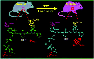
As one of the complications of diabetes, liver injury results in significant hazards. Therefore, accurately diagnosing diabetes-induced liver injury beforehand is crucial for the warning and treatment of hepatic diseases. Diabetes-induced liver injury can cause changes in the microstructure and morphology of liver tissue, leading to changes in the hydrophilic and hydrophobic domains in the endoplasmic reticulum (ER), which is closely associated with changes in cellular ER polarity. So, differences in the ER polarity can indicate the degree of diabetes-induced liver injury. Herein, we develop a new fluorescent and photoacoustic dual-mode probe, ER-P, for detection of the ER polarity of liver tissue in normal and diabetic mice. Upon excitation with a 633 nm laser, ER-P showed increasing fluorescence intensity at 800 nm accompanying a decline in the polarity. Due to its polarity-sensitivity, ER-P was utilized for confocal fluorescence imaging in live cells, and the results demonstrate that ER-P can exclusively accumulate in the ER and indicate an increase in the polarity during ER stress. Importantly, ER-P displayed different absorbance intensities at 700 nm and 800 nm in different polarity environments because of intramolecular charge transfer. The photoacoustic intensity ratios between 700 nm and 800 nm will enable quantification of polarity to be achieved. The ratiometric photoacoustic imaging data demonstrate that the polarity of the liver tissue of diabetic mice is higher than that of the liver tissue of normal mice. Meanwhile, after treatment with the antidiabetic drug metformin, diabetic mice exhibit a reduced polarity environment in their liver tissue. The proposed study may serve as a new approach for the early diagnosis and therapeutic evaluation of diabetes-induced liver injury.
中文翻译:

糖尿病小鼠肝组织内质网极性的比例光声成像
作为糖尿病的并发症之一,肝损伤会导致严重的危害。因此,事先正确诊断糖尿病引起的肝损伤对于肝病的预警和治疗至关重要。糖尿病引起的肝损伤可导致肝组织的微观结构和形态发生变化,从而导致内质网(ER)中亲水和疏水域的变化,这与细胞ER极性的变化密切相关。因此,ER极性的差异可以表明糖尿病引起的肝损伤的程度。本文中,我们开发了一种新型的荧光和光声双模探针ER-P,用于检测正常和糖尿病小鼠肝组织的ER极性。用633 nm激光激发后,ER-P在800 nm处显示出增加的荧光强度,伴随着极性的下降。由于其极性敏感性,ER-P被用于活细胞的共聚焦荧光成像,结果表明ER-P可以仅在ER中积累,并表明在ER应激过程中极性增加。重要的是,由于分子内电荷转移,ER-P在不同极性环境下在700 nm和800 nm处显示出不同的吸收强度。700 nm和800 nm之间的光声强度比将使极性量化成为可能。比例光声成像数据表明,糖尿病小鼠肝组织的极性高于正常小鼠肝组织的极性。同时,在用降糖药二甲双胍治疗后,糖尿病小鼠在其肝组织中显示出极性降低的环境。拟议的研究可作为糖尿病诱发的肝损伤的早期诊断和治疗评估的新方法。
更新日期:2017-09-25
中文翻译:
糖尿病小鼠肝组织内质网极性的比例光声成像
作为糖尿病的并发症之一,肝损伤会导致严重的危害。因此,事先正确诊断糖尿病引起的肝损伤对于肝病的预警和治疗至关重要。糖尿病引起的肝损伤可导致肝组织的微观结构和形态发生变化,从而导致内质网(ER)中亲水和疏水域的变化,这与细胞ER极性的变化密切相关。因此,ER极性的差异可以表明糖尿病引起的肝损伤的程度。本文中,我们开发了一种新型的荧光和光声双模探针ER-P,用于检测正常和糖尿病小鼠肝组织的ER极性。用633 nm激光激发后,ER-P在800 nm处显示出增加的荧光强度,伴随着极性的下降。由于其极性敏感性,ER-P被用于活细胞的共聚焦荧光成像,结果表明ER-P可以仅在ER中积累,并表明在ER应激过程中极性增加。重要的是,由于分子内电荷转移,ER-P在不同极性环境下在700 nm和800 nm处显示出不同的吸收强度。700 nm和800 nm之间的光声强度比将使极性量化成为可能。比例光声成像数据表明,糖尿病小鼠肝组织的极性高于正常小鼠肝组织的极性。同时,在用降糖药二甲双胍治疗后,糖尿病小鼠在其肝组织中显示出极性降低的环境。拟议的研究可作为糖尿病诱发的肝损伤的早期诊断和治疗评估的新方法。


 京公网安备 11010802027423号