Advanced Drug Delivery Reviews ( IF 16.1 ) Pub Date : 2017-07-19 , DOI: 10.1016/j.addr.2017.07.012 Xiang Li , Marcus Hacker
|
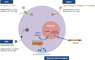
In the past 15 years, despite that regenerative medicine has shown great potential for cardiovascular diseases, the outcome and safety of stem cell transplantation has shown controversial results in the published literature. Medical imaging might be useful for monitoring and quantifying transplanted cells within the heart and to serially characterize the effects of stem cell therapy of the myocardium. From the multiple available noninvasive imaging techniques, magnetic resonance imaging and nuclear imaging by positron (PET) or single photon emission computer tomography (SPECT) are the most used clinical approaches to follow the fate of transplanted stem cells in vivo. In this article, we provide a review on the role of different noninvasive imaging modalities and discuss their advantages and disadvantages. We focus on the different in-vivo labeling and reporter gene imaging strategies for stem cell tracking as well as the concept and reliability to use imaging parameters as noninvasive surrogate endpoints for the evaluation of the post-therapeutic outcome.
中文翻译:
基于干细胞的心脏病治疗中的分子成像
在过去的15年中,尽管再生医学在心血管疾病中显示出巨大的潜力,但干细胞移植的结果和安全性在已发表的文献中显示了有争议的结果。医学成像可能对监视和量化心脏内的移植细胞以及连续表征心肌干细胞治疗的效果很有用。从多种可用的非侵入性成像技术中,通过正电子(PET)或单光子发射计算机断层扫描(SPECT)进行磁共振成像和核成像是追踪体内干细胞命运的最常用临床方法。在本文中,我们对不同的非侵入性成像方式的作用进行了综述,并讨论了它们的优缺点。我们专注于干细胞追踪的不同体内标记和报告基因成像策略,以及使用成像参数作为非侵入性替代终点评估治疗后结果的概念和可靠性。


 京公网安备 11010802027423号