当前位置:
X-MOL 学术
›
Biomaterials
›
论文详情
Our official English website, www.x-mol.net, welcomes your feedback! (Note: you will need to create a separate account there.)
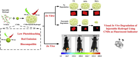
Visual in vivo degradation of injectable hydrogel by real-time and non-invasive tracking using carbon nanodots as fluorescent indicator
Biomaterials ( IF 14.0 ) Pub Date : 2017-08-26 , DOI: 10.1016/j.biomaterials.2017.08.039 Lei Wang , Baoqiang Li , Feng Xu , Ying Li , Zheheng Xu , Daqing Wei , Yujie Feng , Yaming Wang , Dechang Jia , Yu Zhou
Biomaterials ( IF 14.0 ) Pub Date : 2017-08-26 , DOI: 10.1016/j.biomaterials.2017.08.039 Lei Wang , Baoqiang Li , Feng Xu , Ying Li , Zheheng Xu , Daqing Wei , Yujie Feng , Yaming Wang , Dechang Jia , Yu Zhou
|
Visual in vivo degradation of hydrogel by fluorescence-related tracking and monitoring is crucial for quantitatively depicting the degradation profile of hydrogel in a real-time and non-invasive manner. However, the commonly used fluorescent imaging usually encounters limitations, such as intrinsic photobleaching of organic fluorophores and uncertain perturbation of degradation induced by the change in molecular structure of hydrogel. To address these problems, we employed photoluminescent carbon nanodots (CNDs) with low photobleaching, red emission and good biocompatibility as fluorescent indicator for real-time and non-invasive visual in vitro/in vivo degradation of injectable hydrogels that are mixed with CNDs. The in vitro/in vivo toxicity results suggested that CNDs were nontoxic. The embedded CNDs in hydrogels did not diffuse outside in the absence of hydrogel degradation. We had acquired similar degradation kinetics (PBS-Enzyme) between gravimetric and visual determination, and established mathematical equation to quantitatively depict in vitro degradation profile of hydrogels for the predication of in vivo hydrogel degradation. Based on the in vitro data, we developed a visual platform that could quantitatively depict in vivo degradation behavior of new injectable biomaterials by real-time and non-invasive fluorescence tracking. This fluorescence-related visual imaging methodology could be applied to subcutaneous degradation of injectable hydrogel with down to 7 mm depth in small animal trials so far. This fluorescence-related visual imaging methodology holds great potentials for rational design and convenient in vivo screening of biocompatible and biodegradable injectable hydrogels in tissue engineering.
中文翻译:

使用碳纳米点作为荧光指示剂,通过实时和非侵入式跟踪,可视化体内注射性水凝胶的降解
通过荧光相关的跟踪和监视对水凝胶进行可视的体内降解对于以实时且非侵入性的方式定量描述水凝胶的降解情况至关重要。然而,通常使用的荧光成像通常会遇到局限性,例如有机荧光团的固有光漂白和由水凝胶分子结构的变化引起的降解的不确定扰动。为了解决这些问题,我们采用了具有低光致漂白,红色发射和良好生物相容性的光致发光碳纳米点(CND)作为荧光指示剂,可实时,无创地观察与CND混合的可注射水凝胶的体外/体内降解。在体外/体内毒性结果表明CND是无毒的。在没有水凝胶降解的情况下,水凝胶中嵌入的CND不会扩散到外部。我们已经获得了重量测定法和视觉测定法之间相似的降解动力学(PBS-酶),并建立了数学方程式以定量描述水凝胶的体外降解曲线,以预测体内水凝胶的降解。根据体外数据,我们开发了可以定量描述体内的视觉平台实时和非侵入式荧光跟踪技术对新型可注射生物材料的降解行为。迄今为止,这种与荧光有关的视觉成像方法可用于皮下降解可注射水凝胶的深度不超过7毫米的小型动物试验。这种与荧光有关的视觉成像方法在组织工程中对生物相容性和可生物降解的可注射水凝胶进行合理设计和方便的体内筛选方面具有巨大潜力。
更新日期:2017-08-28
中文翻译:
使用碳纳米点作为荧光指示剂,通过实时和非侵入式跟踪,可视化体内注射性水凝胶的降解
通过荧光相关的跟踪和监视对水凝胶进行可视的体内降解对于以实时且非侵入性的方式定量描述水凝胶的降解情况至关重要。然而,通常使用的荧光成像通常会遇到局限性,例如有机荧光团的固有光漂白和由水凝胶分子结构的变化引起的降解的不确定扰动。为了解决这些问题,我们采用了具有低光致漂白,红色发射和良好生物相容性的光致发光碳纳米点(CND)作为荧光指示剂,可实时,无创地观察与CND混合的可注射水凝胶的体外/体内降解。在体外/体内毒性结果表明CND是无毒的。在没有水凝胶降解的情况下,水凝胶中嵌入的CND不会扩散到外部。我们已经获得了重量测定法和视觉测定法之间相似的降解动力学(PBS-酶),并建立了数学方程式以定量描述水凝胶的体外降解曲线,以预测体内水凝胶的降解。根据体外数据,我们开发了可以定量描述体内的视觉平台实时和非侵入式荧光跟踪技术对新型可注射生物材料的降解行为。迄今为止,这种与荧光有关的视觉成像方法可用于皮下降解可注射水凝胶的深度不超过7毫米的小型动物试验。这种与荧光有关的视觉成像方法在组织工程中对生物相容性和可生物降解的可注射水凝胶进行合理设计和方便的体内筛选方面具有巨大潜力。


 京公网安备 11010802027423号