当前位置:
X-MOL 学术
›
Metallomics
›
论文详情
Our official English website, www.x-mol.net, welcomes your feedback! (Note: you will need to create a separate account there.)
Evidence for widespread, severe brain copper deficiency in Alzheimer's dementia†
Metallomics ( IF 3.4 ) Pub Date : 2017-06-16 00:00:00 , DOI: 10.1039/c7mt00074j Jingshu Xu 1, 2, 3, 4, 5 , Stephanie J. Church 6, 7, 8, 9, 10 , Stefano Patassini 1, 2, 3, 4, 5 , Paul Begley 6, 7, 8, 9, 10 , Henry J. Waldvogel 3, 5, 11, 12, 13 , Maurice A. Curtis 3, 5, 11, 12, 13 , Richard L. M. Faull 3, 5, 11, 12, 13 , Richard D. Unwin 6, 7, 8, 9, 10 , Garth J. S. Cooper 1, 2, 3, 4, 5
Metallomics ( IF 3.4 ) Pub Date : 2017-06-16 00:00:00 , DOI: 10.1039/c7mt00074j Jingshu Xu 1, 2, 3, 4, 5 , Stephanie J. Church 6, 7, 8, 9, 10 , Stefano Patassini 1, 2, 3, 4, 5 , Paul Begley 6, 7, 8, 9, 10 , Henry J. Waldvogel 3, 5, 11, 12, 13 , Maurice A. Curtis 3, 5, 11, 12, 13 , Richard L. M. Faull 3, 5, 11, 12, 13 , Richard D. Unwin 6, 7, 8, 9, 10 , Garth J. S. Cooper 1, 2, 3, 4, 5
Affiliation

|
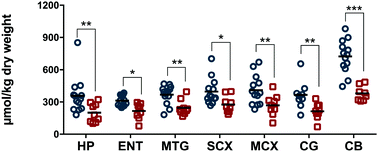
Datasets comprising simultaneous measurements of many essential metals in Alzheimer's disease (AD) brain are sparse, and available studies are not entirely in agreement. To further elucidate this matter, we employed inductively-coupled-plasma mass spectrometry to measure post-mortem levels of 8 essential metals and selenium, in 7 brain regions from 9 cases with AD (neuropathological severity Braak IV–VI), and 13 controls who had normal ante-mortem mental function and no evidence of brain disease. Of the regions studied, three undergo severe neuronal damage in AD (hippocampus, entorhinal cortex and middle-temporal gyrus); three are less-severely affected (sensory cortex, motor cortex and cingulate gyrus); and one (cerebellum) is relatively spared. Metal concentrations in the controls differed among brain regions, and AD-associated perturbations in most metals occurred in only a few: regions more severely affected by neurodegeneration generally showed alterations in more metals, and cerebellum displayed a distinctive pattern. By contrast, copper levels were substantively decreased in all AD-brain regions, to 52.8–70.2% of corresponding control values, consistent with pan-cerebral copper deficiency. This copper deficiency could be pathogenic in AD, since levels are lowered to values approximating those in Menkes’ disease, an X-linked recessive disorder where brain-copper deficiency is the accepted cause of severe brain damage. Our study reinforces others reporting deficient brain copper in AD, and indicates that interventions aimed at safely and effectively elevating brain copper could provide a new experimental-therapeutic approach.
中文翻译:

阿尔茨海默氏痴呆症广泛,严重的脑铜缺乏的证据†
包括同时测量阿尔茨海默氏病(AD)大脑中许多必需金属的数据集稀疏,可用的研究并不完全一致。为了进一步阐明此问题,我们采用了电感耦合等离子体质谱法来进行验尸9例AD(神经病理学严重程度为Braak IV-VI)的7个大脑区域和13个具有正常事前验血功能且无脑疾病证据的对照中8种必需金属和硒的水平。在研究的区域中,三个区域在AD中遭受了严重的神经元损伤(海马,内嗅皮层和中颞回)。三种受影响程度较轻(感觉皮层,运动皮层和扣带回);而一个(小脑)则相对幸免。对照区域中的金属浓度在大脑区域之间有所不同,大多数金属中与AD相关的扰动仅在少数情况下发生:受神经变性影响更严重的区域通常显示更多金属的变化,而小脑表现出独特的模式。相比之下,在所有AD脑区域中,铜含量均显着下降,降至52.8–70。相应对照值的2%,与全脑铜缺乏症一致。这种铜缺乏症在AD中可能是致病的,因为其水平降低至接近Menkes病的水平,Menkes病是一种X连锁隐性疾病,其中脑铜缺乏是导致严重脑损伤的公认原因。我们的研究强化了其他报道AD中脑铜缺乏的人,并表明旨在安全有效地升高脑铜的干预措施可以提供一种新的实验治疗方法。
更新日期:2017-06-16
中文翻译:

阿尔茨海默氏痴呆症广泛,严重的脑铜缺乏的证据†
包括同时测量阿尔茨海默氏病(AD)大脑中许多必需金属的数据集稀疏,可用的研究并不完全一致。为了进一步阐明此问题,我们采用了电感耦合等离子体质谱法来进行验尸9例AD(神经病理学严重程度为Braak IV-VI)的7个大脑区域和13个具有正常事前验血功能且无脑疾病证据的对照中8种必需金属和硒的水平。在研究的区域中,三个区域在AD中遭受了严重的神经元损伤(海马,内嗅皮层和中颞回)。三种受影响程度较轻(感觉皮层,运动皮层和扣带回);而一个(小脑)则相对幸免。对照区域中的金属浓度在大脑区域之间有所不同,大多数金属中与AD相关的扰动仅在少数情况下发生:受神经变性影响更严重的区域通常显示更多金属的变化,而小脑表现出独特的模式。相比之下,在所有AD脑区域中,铜含量均显着下降,降至52.8–70。相应对照值的2%,与全脑铜缺乏症一致。这种铜缺乏症在AD中可能是致病的,因为其水平降低至接近Menkes病的水平,Menkes病是一种X连锁隐性疾病,其中脑铜缺乏是导致严重脑损伤的公认原因。我们的研究强化了其他报道AD中脑铜缺乏的人,并表明旨在安全有效地升高脑铜的干预措施可以提供一种新的实验治疗方法。


 京公网安备 11010802027423号