Our official English website, www.x-mol.net, welcomes your feedback! (Note: you will need to create a separate account there.)
Trace‐Element Incorporation into Intracellular Pools Uncovers Calcium‐Pathways in a Coccolithophore
Advanced Science ( IF 15.1 ) Pub Date : 2017-07-05 , DOI: 10.1002/advs.201700088 Assaf Gal 1, 2 , Sanja Sviben 1 , Richard Wirth 3 , Anja Schreiber 3 , Benedikt Lassalle-Kaiser 4 , Damien Faivre 2 , André Scheffel 1
Advanced Science ( IF 15.1 ) Pub Date : 2017-07-05 , DOI: 10.1002/advs.201700088 Assaf Gal 1, 2 , Sanja Sviben 1 , Richard Wirth 3 , Anja Schreiber 3 , Benedikt Lassalle-Kaiser 4 , Damien Faivre 2 , André Scheffel 1
Affiliation
|
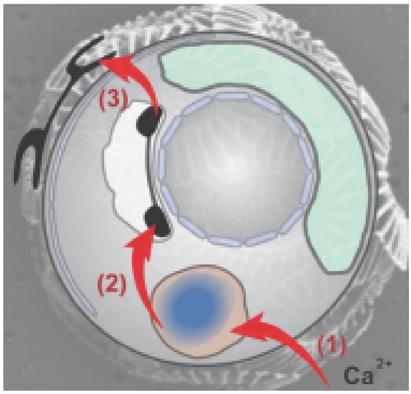
Many organisms form minerals from precursor phases that crystallize under strict biological control. The dynamic intracellular processes of formation, transport, and deposition of these precursor phases are challenging to identify. An unusual situation is recently revealed for the calcifying alga Emiliania huxleyi, as the cells contain a compartment filled with a concentrated Ca and P phase but the final calcite crystals, which are nucleated in a different compartment, are P‐free. Thus, the connection of the Ca–P‐rich pool to the mineralization process remains unclear. Here, pulse‐chase experiments are used with Sr to label the Ca–P‐rich phase in E. huxleyi cells, and cryo X‐ray absorption spectroscopy and analytical transmission electron microscopy to follow the Sr within cells. It is found that Sr is first found in the Ca–P‐rich phase and then becomes incorporated into the calcite. This demonstrates that the calcium used by the cells to build calcite originates from the Ca–P‐rich pool.
中文翻译:

痕量元素掺入细胞内池发现了Coccolithophore中的钙途径。
许多生物从前体相中形成矿物质,这些前体相在严格的生物控制下会结晶。这些前体相的形成,运输和沉积的动态细胞内过程难以确定。最近发现钙化藻类Emiliania huxleyi出现了一种不寻常的情况,因为细胞包含一个充满浓缩的Ca和P相的隔室,但是在不同隔室中成核的最终方解石晶体不含P。因此,尚不清楚钙富集池与成矿过程的联系。在这里,脉冲追踪实验与Sr一起使用以标记赫氏大肠杆菌中的富含Ca-P的相细胞,冷冻X射线吸收光谱法和分析型透射电子显微镜观察细胞内的Sr. 已经发现,Sr首先在富Ca-P相中发现,然后被掺入方解石中。这表明细胞用来构建方解石的钙起源于富含Ca-P的池。
更新日期:2017-07-05
中文翻译:
痕量元素掺入细胞内池发现了Coccolithophore中的钙途径。
许多生物从前体相中形成矿物质,这些前体相在严格的生物控制下会结晶。这些前体相的形成,运输和沉积的动态细胞内过程难以确定。最近发现钙化藻类Emiliania huxleyi出现了一种不寻常的情况,因为细胞包含一个充满浓缩的Ca和P相的隔室,但是在不同隔室中成核的最终方解石晶体不含P。因此,尚不清楚钙富集池与成矿过程的联系。在这里,脉冲追踪实验与Sr一起使用以标记赫氏大肠杆菌中的富含Ca-P的相细胞,冷冻X射线吸收光谱法和分析型透射电子显微镜观察细胞内的Sr. 已经发现,Sr首先在富Ca-P相中发现,然后被掺入方解石中。这表明细胞用来构建方解石的钙起源于富含Ca-P的池。


 京公网安备 11010802027423号