当前位置:
X-MOL 学术
›
J. Am. Chem. Soc.
›
论文详情
Our official English website, www.x-mol.net, welcomes your feedback! (Note: you will need to create a separate account there.)
Ultrafast Elemental and Oxidation-State Mapping of Hematite by 4D Electron Microscopy
Journal of the American Chemical Society ( IF 15.0 ) Pub Date : 2017-03-21 , DOI: 10.1021/jacs.7b00906 Zixue Su 1 , J. Spencer Baskin 1 , Wuzong Zhou 2 , John M. Thomas 3 , Ahmed H. Zewail 1
Journal of the American Chemical Society ( IF 15.0 ) Pub Date : 2017-03-21 , DOI: 10.1021/jacs.7b00906 Zixue Su 1 , J. Spencer Baskin 1 , Wuzong Zhou 2 , John M. Thomas 3 , Ahmed H. Zewail 1
Affiliation

|
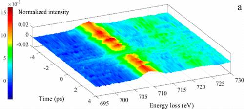
We describe a new methodology that sheds light on the fundamental electronic processes that occur at the subsurface regions of inorganic solid photocatalysts. Three distinct kinds of microscopic imaging are used that yield spatial, temporal, and energy-resolved information. We also carefully consider the effect of photon-induced near-field electron microscopy (PINEM), first reported by Zewail et al. in 2009. The value of this methodology is illustrated by studying afresh a popular and viable photocatalyst, hematite, α-Fe2O3 that exhibits most of the properties required in a practical application. By employing high-energy electron-loss signals (of several hundred eV), coupled to femtosecond temporal resolution as well as ultrafast energy-filtered transmission electron microscopy in 4D, we have, inter alia, identified Fe4+ ions that have a lifetime of a few picoseconds, as well as associated photoinduced electronic transitions and charge transfer processes.
中文翻译:

通过 4D 电子显微镜对赤铁矿进行超快元素和氧化状态映射
我们描述了一种新方法,它阐明了发生在无机固体光催化剂的亚表面区域的基本电子过程。使用三种不同的显微成像来产生空间、时间和能量分辨信息。我们还仔细考虑了 Zewail 等人首先报道的光子诱导近场电子显微镜 (PINEM) 的影响。2009 年。通过重新研究一种流行且可行的光催化剂,赤铁矿,α-Fe2O3,展示了实际应用中所需的大部分特性,说明了该方法的价值。通过采用高能电子损耗信号(数百 eV),结合飞秒时间分辨率以及 4D 中的超快能量过滤透射电子显微镜,我们除其他外,
更新日期:2017-03-21
中文翻译:

通过 4D 电子显微镜对赤铁矿进行超快元素和氧化状态映射
我们描述了一种新方法,它阐明了发生在无机固体光催化剂的亚表面区域的基本电子过程。使用三种不同的显微成像来产生空间、时间和能量分辨信息。我们还仔细考虑了 Zewail 等人首先报道的光子诱导近场电子显微镜 (PINEM) 的影响。2009 年。通过重新研究一种流行且可行的光催化剂,赤铁矿,α-Fe2O3,展示了实际应用中所需的大部分特性,说明了该方法的价值。通过采用高能电子损耗信号(数百 eV),结合飞秒时间分辨率以及 4D 中的超快能量过滤透射电子显微镜,我们除其他外,


 京公网安备 11010802027423号