当前位置:
X-MOL 学术
›
Chem. Rev.
›
论文详情
Our official English website, www.x-mol.net, welcomes your feedback! (Note: you will need to create a separate account there.)
Single-Molecule Localization Microscopy in Eukaryotes
Chemical Reviews ( IF 62.1 ) Pub Date : 2017-03-13 00:00:00 , DOI: 10.1021/acs.chemrev.6b00667 Markus Sauer 1 , Mike Heilemann 2
Chemical Reviews ( IF 62.1 ) Pub Date : 2017-03-13 00:00:00 , DOI: 10.1021/acs.chemrev.6b00667 Markus Sauer 1 , Mike Heilemann 2
Affiliation
|
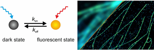
Super-resolution fluorescence imaging by photoactivation or photoswitching of single fluorophores and position determination (single-molecule localization microscopy, SMLM) provides microscopic images with subdiffraction spatial resolution. This technology has enabled new insights into how proteins are organized in a cellular context, with a spatial resolution approaching virtually the molecular level. A unique strength of SMLM is that it delivers molecule-resolved information, along with super-resolved images of cellular structures. This allows quantitative access to cellular structures, for example, how proteins are distributed and organized and how they interact with other biomolecules. Ultimately, it is even possible to determine protein numbers in cells and the number of subunits in a protein complex. SMLM thus has the potential to pave the way toward a better understanding of how cells function at the molecular level. In this review, we describe how SMLM has contributed new knowledge in eukaryotic biology, and we specifically focus on quantitative biological data extracted from SMLM images.
中文翻译:

真核生物中的单分子定位显微镜
通过单个荧光团的光激活或光转换以及位置确定(单分子定位显微镜,SMLM)进行的超分辨率荧光成像可提供具有亚衍射空间分辨率的显微图像。这项技术使人们对蛋白质在细胞环境中的组织方式有了新的认识,而空间分辨率实际上接近了分子水平。SMLM的独特优势在于,它可以提供分子解析的信息以及细胞结构的超解析图像。这允许定量访问细胞结构,例如蛋白质如何分布和组织以及它们如何与其他生物分子相互作用。最终,甚至有可能确定细胞中的蛋白质数量和蛋白质复合物中的亚基数量。因此,SMLM有可能为更好地了解细胞在分子水平上的功能铺平道路。在这篇综述中,我们描述了SMLM如何为真核生物学贡献了新知识,并且我们特别关注从SMLM图像中提取的定量生物学数据。
更新日期:2017-03-13
中文翻译:
真核生物中的单分子定位显微镜
通过单个荧光团的光激活或光转换以及位置确定(单分子定位显微镜,SMLM)进行的超分辨率荧光成像可提供具有亚衍射空间分辨率的显微图像。这项技术使人们对蛋白质在细胞环境中的组织方式有了新的认识,而空间分辨率实际上接近了分子水平。SMLM的独特优势在于,它可以提供分子解析的信息以及细胞结构的超解析图像。这允许定量访问细胞结构,例如蛋白质如何分布和组织以及它们如何与其他生物分子相互作用。最终,甚至有可能确定细胞中的蛋白质数量和蛋白质复合物中的亚基数量。因此,SMLM有可能为更好地了解细胞在分子水平上的功能铺平道路。在这篇综述中,我们描述了SMLM如何为真核生物学贡献了新知识,并且我们特别关注从SMLM图像中提取的定量生物学数据。


 京公网安备 11010802027423号